The IX53 has been designed from the ground up to be the finest microscope available for routine inverted microscopic analysis. The combination of excellent optical performance and mechanical quality results in a microscope system of outstanding value and comfort for regular use.
Features such as the pre-centered phase contrast, relief contrast and the flexible UIS2 DIC optics enable easy visualization across all magnifications for both thin and thick specimens. Available condensers with different working distances and long working objectives allow observation and documentation of even the most complex samples.
Features
Expandable to Meet Growing Research Needs
The IX53 system is the ideal solution for routine analysis. End-users can improve research efficiency by recording cell dynamics across a wide area. Reliability can also be enhanced through accurate position and light reproducibility.
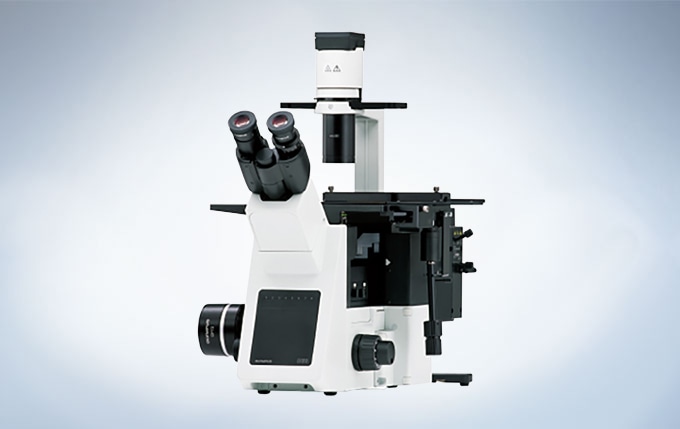
IX53: One-deck System
An outstanding, cost-effective microscope for brightfield and fluorescence applications.
Reliable, Clear and High-Resolution Images
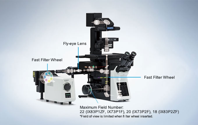
Evident UIS2 infinity-corrected optics ensure high optical transmittance with a broad range of objectives. UIS2 optics feature wide chromatic correction and enable high resolution, high S/N primary images regardless of the observation method. The wide field of view and Fly-Eye lens system provide uniform fluorescence images and enable the use of sCMOS cameras with large sensors.
Excellent Image Quality
Apochromatic Objectives Enable High-Resolution Phase Contrast and Fluorescence Observation
Phase contrast apochromatic objectives (UPLSAPO100XOPH, PLAPON60XOPH) enable high-precision imaging free from image shift, even during simultaneous phase contrast and fluorescence observation. This eliminates the need to change objectives when switching observation methods.
HeLa cell expressing mCherry-actin (Image data courtesy of: Tomonobu Watanabe, Ph.D. Laboratory for Comprehensive Bioimaging, RIKEN Quantitative Biology Center)
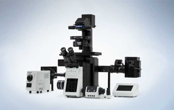
IX83:Two-deck System
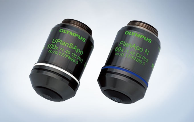
UPLSAPO100×OPH, PLAPON60×OPH
Silicone Objectives* Enable High-Resolution Observation Deep into Live Cells
Evident offers three high-NA silicone immersion objectives:UPLSAPO30XS, UPLSAPO40XS, and UPLSAPO60XS. The refractive index of silicone oil (Refractive index: ne≈1.40) is close to that of living tissue (Refractive index: ne≈1.38), enabling high-resolution observation deep inside living tissue with minimal spherical aberration caused by refractive index mismatch. Silicone oil does not dry out or harden, so there is never a need to readminister the oil, making it ideal for extended time-lapse observations.
*Use dedicated silicone oil.
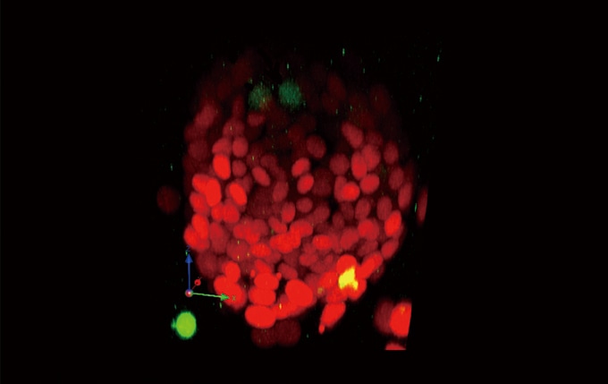
3D reconstruction images of a live sphere made of NMuMG/Fucci2 cells. Confocal images were acquired by using an Evident FV1000 confocal microscope. (Red: cell cycle G1 phase, Green: cell cycle S/G2/M phase) Image data courtesy of: Asako Sakaue-Sawano, Ph.D. Atsushi Miyawaki, M.D., Ph.D. Laboratory for Cell Function Dynamics, Advanced Technology Development Core, RIKEN Brain Science Institute
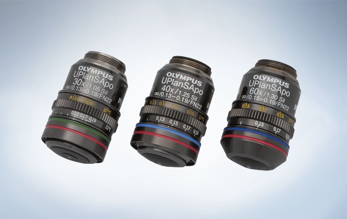
UPLSAPO30XS, UPLSAPO40XS and UPLSAPO60XS
Special Objective Available for iPS/ES and Floating Cell Observation
This high-NA phase contrast objective (UCPLFLN20XPH) is especially suited for observation using plastic dishes. It enables high-resolution observation of the cell proliferation process and delivers improved contrast across a wide area.
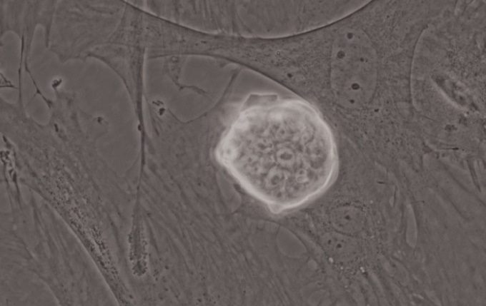
iPS-cell expressing Nanog reporter (GFP) Image data courtesy of: Tomonobu Watanabe, Ph.D. Laboratory for Comprehensive Bioimaging, RIKEN Quantitative Biology Center
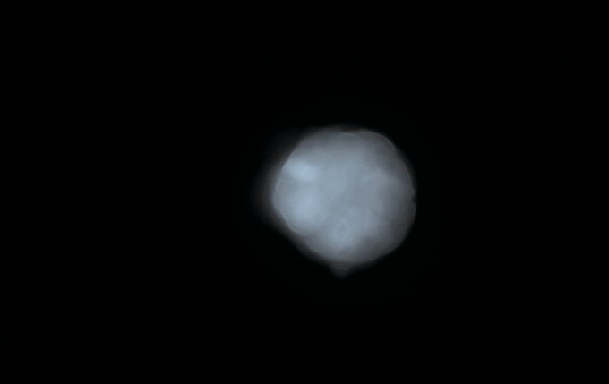
iPS-cell expressing Nanog reporter (GFP) Image data courtesy of: Tomonobu Watanabe, Ph.D. Laboratory for Comprehensive Bioimaging, RIKEN Quantitative Biology Center
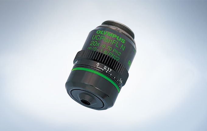
UCPLFLN20×PH
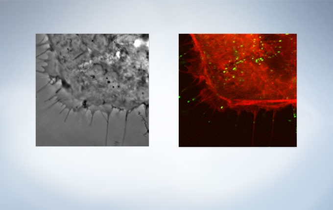
High S/N Fluorescence Mirror Units for Efficient Signal Detection
All fluorescence mirror units feature filters treated with a specially-developed coating that minimizes noise by absorbing more than 99 % of stray light. The outstanding performance and high transmittance of the mirror units ensure efficient fluorescence signal detection.
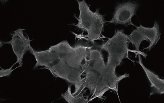
Image Captured with a Conventional FL Mirror Unit

Images Captured with New FL Mirror Units
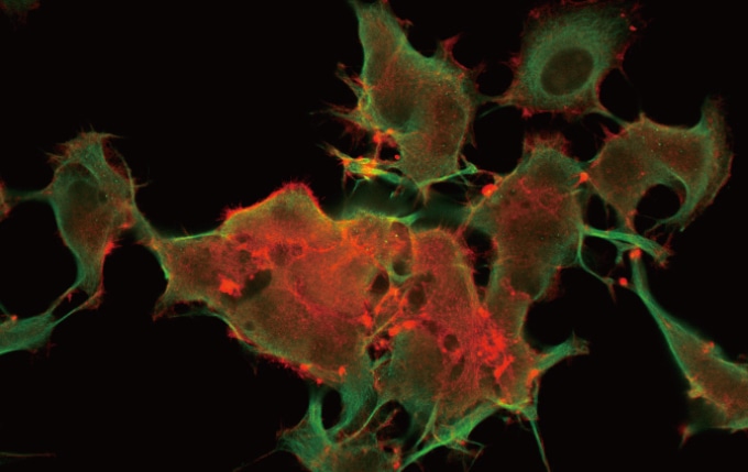
Images Captured with New FL Mirror Units