Monoclonal antibodies (mAbs) represent one of the fastest growing classes of pharmaceuticals, and look set to be the focus of the biotherapeutics industry into the years to come. These proteins possess enormous potential from a drug development point-of-view as they can be engineered to recognise almost any antigen and can subsequently be mass-produced in large quantities.
owever, the correct folding of mAbs is critical for their efficacy, as their structure is closely related to their function. Incorrect folding can impact safety, for instance, by inducing an unwanted immune response. Therefore, methods to quickly and accurately characterise the structure of mAbs are needed for quality control purposes.
Making progress
The high-resolution structural analysis of mAbs has largely been thought to be unfeasible due to the limitations of available techniques, such as X-ray crystallography and nuclear magnetic resonance (NMR) spectroscopy.
For example, the size of mAbs, at around 150 kDa, decreases the quality of the spectra produced by NMR and their nature also results in line broadening, reducing the sensitivity of the results.
Furthermore, due to the low natural abundance of the isotopes 13C and 15N (1.1 and 0.37%, respectively), 2D NMR of biomolecules usually requires isotopic labelling, which is not readily available in a quality control setting.
However, advances in the field of spectroscopy are beginning to overcome these challenges. For example, the emergence of cryogenic probe technology is making 2D NMR spectroscopy at natural isotopic abundance much more feasible, as is the greater accessibility of high spectrometer field strengths over recent years.
Furthermore, newly developed acquisition methods, such as the SOFAST (selective optimised flip angle short transient) pulse scheme, have reduced the time needed for natural abundance experiments, particularly when combined with non-uniform sampling methods.
In a recent study, researchers led by Luke Arbogast explored how these advances could allow rapid and detailed characterisation of mAbs.
A new approach
In the study, the researchers used 2D NMR to analyse a candidate mAb called RM8670. They chose to look at the methyl resonances of the protein to take advantage of the greater natural abundance of the 13C isotope over 15N.
This contrasts the more established approach of mapping the nitrogen-rich amide backbone of the protein. Methyl groups are well distributed throughout proteins, the researchers explain and, as they are affected by the hydrophobic packing of the protein, are also good reporters of its 3D structure.
Arbogast et al first performed their experiments using 2D 13C NMR at 900 MHz on samples of intact mAb, using a sequence known as gsHSQC (gradient-selected, sensitivity-enhanced heteronuclear single quantum coherence). They found that this gave “better than expected” resolution and they were able to perform assignment and analysis of the spectrum peaks. However, when they re-conducted the experiments, optimising them to produce the desired spectral quality, the experiment took 12 hours.
To examine whether the experimental time could be reduced, Arbogast et al repeated their experiments using mAb that had been enzymatically cleaved into its Fab and Fc domains. They found that in this experiment, which took only 4.5 hours, the spectrum produced matched closely that produced with intact mAb. This suggests that cleavage does not disrupt the structure of the Fab and Fc fragments and that the Fab/Fc mixture acts as a good proxy for the intact protein.
The researchers then went on to see if experimental times could be reduced even further. They did this by combining rapid acquisition techniques with non-uniform sampling and applying them to Fc and Fab fragments. The quickest time they were able to achieve was 34 minutes with a combination of the rapid acquisition technique SOFAST-HMQC with 50% non-uniform sampling.
Writing in Analytical Chemistry, the researchers say their findings demonstrate the potential of natural abundance 2D NMR fingerprinting for the structural assessment of mAbs in situations where isotope labelling is not possible.
NMR provides advantages over other forms of structural assessment in giving a combined readout of primary, secondary, tertiary and higher order structure of a protein at atomic resolution, they write. Their newly developed approach should therefore have wide uses in the biotherapeutic industry for protein structure assessment, the team concludes.
An advance with Avance
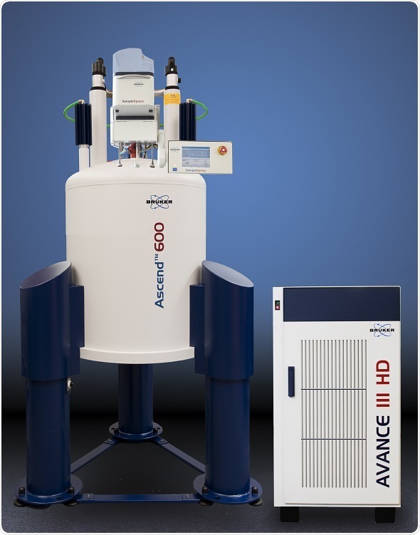
In their experiments, Arbogast et al used Bruker Avance III 900 and 600 MHz spectrometers equipped with triple-resonance cryoprobes.
The Bruker Avance III HD offers fast, flexible NMR with the most advanced RF generation in the industry. Meanwhile, Bruker offers two lines of cryogenic probe technologies that can help assist natural isotopic abundance NMR: CryoProbe, which is based on a closed cycle helium cryocooler and CryoProbe Prodigy which is an open cycle liquid nitrogen cooling system. Both use a range of automated features including cool-down, warm-up and timer, and are compatible with Bruker’s automatic sample changer.
References
- Arbogast LW, Brinson RG & Marino JP. Mapping. Mapping Monoclonal Antibody Structure by 2D 13C NMR at Natural Abundance. Analytical Chemistry 2015; 87: 3556-3561.
- Goswami S, Wang W, Arakawa T, et al. Developments and Challenges for mAb-Based Therapeutics. Antibodies 2013; 2: 452-500.
- Poppe L, Jordan JB, Rogers G, et al. On the Analytical Superiority of 1D NMR for Fingerprinting the Higher Order Structure of Protein Therapeutics Compared to Multidimensional NMR Methods. Anal Chem 2015; 87: 5539-5545.
About Bruker BioSpin - NMR, EPR and Imaging

Bruker BioSpin offers the world's most comprehensive range of NMR and EPR spectroscopy and preclinical research tools. Bruker BioSpin develops, manufactures and supplies technology to research establishments, commercial enterprises and multi-national corporations across countless industries and fields of expertise.
Sponsored Content Policy: News-Medical.net publishes articles and related content that may be derived from sources where we have existing commercial relationships, provided such content adds value to the core editorial ethos of News-Medical.Net which is to educate and inform site visitors interested in medical research, science, medical devices and treatments.