In fluorescence microscopy, a specimen emits energy when activated by light of a certain wavelength. Some substances such as chlorophyll and some minerals will do this naturally, while others will need the help of other chemical triggers.
Fluorescence microscope components
A fluorescence microscope is typically made up of the following components:
- A light source, which may be a xenon or mercury burner, an LED, or a laser.
- An excitation filter, which selects from the light source only the excitation wavelengths that are able to excite the specimen.
- A dichroic beam splitter or mirror transmits and reflects light as a function of wavelength.
- An emission filter, which ensures only light emitted from the sample is transmitted, by selecting for the light of a distinct wavelength.
- A CCD camera, which is the most widely used device for detecting the emitted light. It is usually linked to a computer screen which displays the images generated

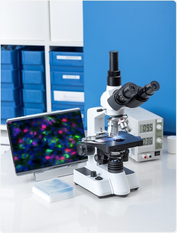
Modern microscope station with antibody-stained tissue sample image on the screen. Image Copyright: anyaivanova / Shutterstock.com
Types of fluorescence microscopy
Epifluorescence microscopy
The Greek word “epi” means the same. In epifluorescence microscopy, the illuminated and emitted light both travel through the same objective lens.
The light source is often a xenon or mercury burner, but more recently, LEDs have become popular. A filter is used to select wavelengths coming from the light source.
The light reaches the specimen via a microscope objective lens and is absorbed by the fluorophores. The emitted light from the sample travels back through the objective lens, towards the emission filter and detector.
Total internal reflection fluorescence (TIRF)
This technique ensures that the excitation light does not make it very far into the sample. For example, if neurons are placed in a solution on a glass slide, some of them will stick to the surface of the glass.
In TIRF, the light is sent sideways into the slide, which means it does not get very far into the solution containing the specimen; it just leaks into the solution, but only very near to the glass surface.
Light is then only emitted in a very thin area against the surface of the glass. This can provide very clear images for a sample such as neurons, where a lot happens on the cell surface.
Confocal fluorescence
Here, a laser spot is focused in the same plane as the focus of the microscope. The light that is not from the microscope focus is blocked by a pinhole in front of the detector.
Ordinarily, with epifluorescence, the microscope would collect all light within the microscope field of view, including light that is not from the microscope focus. This process means the out-of-focus light is blocked and does not reach the detector.
Multiphoton fluorescence
The arrangement of the objective, collection optics, and pinhole in confocal microscopy has to be very precise for the microscope to work properly. Multiphoton fluorescence uses laser light of a wavelength that only has half the energy it needs to be in order to excite the sample.
Fluorophores in the sample will only become excited and emit light when the laser is bright enough for photons to reach the fluorophores very quickly, which only happens when the laser light is directed to a very small spot. Therefore, light is only emitted from the area of the sample where the laser is focused. This eradication of background light results in very clean, sharp images.
Super-resolution fluorescence
Structured illumination microscopy (SIM) and stimulated emission depletion microscopy both work by restricting the size of the spot that emits light by reducing the size of the excitation spot. This is because a focused spot of light can only not get any smaller than around 200nm and single molecules are much smaller than this.
References
Further Reading