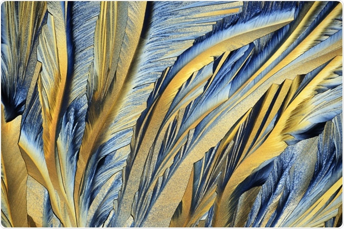
In polarized light microscopy, plane-polarized light is passed through a double refracting material and then collected using a second polarizing filter to generate a high-contrast image. This technique finds application in several fields, such as medicine, basic biology, industry, and to study rock minerals.

Photo through a microscope of crystals growing from the melt of sulfur. Polarized light technology. Image Credit: alex7370 / Shutterstock.com
Medical Applications
Gout diagnosis
Gout occurs because of the deposition of urate crystals in the synovial fluid of the joints which causes inflammation and pain. A polarized microscope is used to examine synovial fluid for the diagnosis of gout.
Urate crystals causing gout have negative elongated optical features, while pyrophosphoric acids which cause pseudo-gout have positive optical features. These differences in the crystal direction and the interference color on polarized light microscopy are used to differentiate these crystals.
Amyloid protein examination
Amyloid proteins are created by the abnormal metabolism of proteins which leads to their aggregation within cells. These proteins may be deposited in various organs, such as the spleen, liver, kidney, brain among others. These aggregates are not observed in normal cells. Using polarized light, these amyloid structures can be visualized. The presence of amyloid proteins is ascertained by the appearance of bright green color on polarization light microscopy.
Applications in Basic Biology
Polarized light microscopy is used to visualize several birefringent or double-refractive structures in the body, including teeth, striated bone, muscle tissue, neurons, spindles, and actomyosin fibers. These structures can be visualized with great contrast by adding a dye; however, as these are living structures, this step causes cell death.
Thus, this technique offers a non-invasive method of high-contrast imaging for these tissues and cells. Polarized light microscopy does not require any contrast agent or dye, can be performed in a non-invasive manner, and generates high-contrast images.
polarizing microscope
Examination of Rocks
Polarized light microscopy can be used to examine rock structures and their optical characteristics. This method can also be used to identify minerals inside rocks. Two methods are used for this purpose.
Transmitted polarizing microscopy
In this method, a rock cutter is used to cut a thin slice of rock. One side is polished and the specimen is fixed onto a glass slide using an adhesive. Subsequently, the opposite side is also polished and fixed to a cover glass using balsam.
Immersion method
In this method, the rock is ground to a fine powder and then sprinkled on a glass slide. The powder is surrounded with an immersion liquid and then a coverslip is added. However, in this method, the structure of the rock is destroyed. The advantage is that it does not require the preparation of thin rock sections.
Applications in Industry
Liquid crystals
Liquid crystals are materials with properties in between liquids and solids. Polarizing microscopes are used to detect peculiar optical patterns and phase defects in liquid crystals. They can also be used to determine if a crystal is optically positive or negative. Liquid crystal retardation can also be measured using polarized microscopes.
Macromolecular materials
Spherulites are transparent, double refractive materials of spherical shape, belonging to the class called macromolecular crystals. Polarized microscopes can be used to determine their density and size which in turn determines the strength and transparency.
Food chemicals
Polarized microscopes can be used to determine the properties of emulsions of butter and cream as they possess optical anisotropy. Thus, using this method, any deviations in the emulsion conditions or impurities can be determined.
Glass and ceramics
Polarized microscopy can be used to determine the quality of and defects in glass and ceramics by identifying their color, shape, diffraction index, and crystal impurities.
Metals
Metal inspection for composition, surface impurities, and properties is possible using polarizing microscopy on a polished metal sample.
Further Reading