Complex, heterogenous systems such as those in immunology, neuroscience, and development can be better understood by studying their associated cellular characteristics. This field has been called “cytometry”. Although this is a costly and time-consuming process, recently developed “ghost cytometry” offers quick and reliable methods to morphologically identify cells.

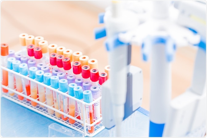
BD Vacutainer tubes, vacuum tubes for collecting blood samples in the lab. Image Credit: science photo / Shutterstock.com
Drawbacks of Previous Methods
Immunophenotyping
Cytometry is an important field of study, with growing interest in cancer research. Analysis of subsets of cells by “immunophenotyping” detects differences in antigens, which can identify cancer cells in leukemia. This is used clinically to detect cancer and subclassify it. However, this method relies on the presence of a biological marker.
Flow cytometry
Examining cell morphology is another method of cell sorting which does not require the presence or discovery of a biological marker to classify cells. However, high quality, usable image construction is difficult and costly due the demands of continuous image acquisition, shutter rate, and frame rate. Secondly, the image then needs to be computationally reconstructed and analyzed, which is both expensive and laborious. Huge amounts of data produced by image flow cytometry (IFC), the widely used method to morphologically assess cancerous cells, are difficult to handle.
Ghost cytometry solves these problems by producing high quality images that can be sorted by machine learning.
Ghost Cytometry - the Principle
Ghost imaging
Ghost cytometry does not not make an image in real-time but combines light emitted from fluorescence and machine learning to classify cells. Traditional “ghost imaging” is a technique by which an object is indirectly visualized by light where an image is indirectly produced by the correlations between two light beams.
The object of interest is moved across a static optical structure. Fluorophores in the object of interest are thus excited, and a single pixel detector measures the intensities of the fluorophores as one combined temporal waveform. The temporal waveform that is produced is the result of an interaction between the intensity distribution of the optical structure and of the moving object.
Image reconstruction
Like the name, the image reconstruction in ghost cytometry shares roots with ghost imaging. Once the single pixel detector has absorbed the signals from the optical pattern, the image is computationally recovered using a special algorithm. The image reconstruction in ghost cytometry is up to 10,000 times faster than in ghost imaging.
Machine Learning
Handling large data
One of the defining factors of ghost cytometry is that the images are analyzed in real time. While new technologies allow data to be collected at high accuracy, the sheer volume of information can be difficult to handle. To retrieve usable information from the ghost cytometry image, Ota and colleagues used machine learning.
Support vector machine
To avoid massive amounts of data, only the waveform data was kept and not necessarily transformed into an image. This method works for machine learning models. The type of machine learning used is called “support vector machine” (SVM), where the algorithm is shown examples of each cell type and then it constructs a model that can distinguish cells into one of those two cell types.
In ghost cytometry, this enables real-time classification of cells by computer based on the wavelengths emitted. This removes the reliance on human competence, massively expanding the potential use of ghost cytometry.
Applications
To ensure their ghost cytometry method was viable, Ota and colleagues tested the method on pancreatic cancer cells. The ghost cytometer could distinguish between cancerous and non-cancerous cells, even when these were similar in size, fluorescent intensity, and morphological features.
The method proved accurate when the cell types were present in blood and in different concentrations. Thus, it has a high level of precision, given that the classification of very similar cell types has previously been an issue for cytometers and human analyzers when biological markers are lacking.
Further Reading