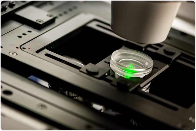
Two-photon, or multiphoton microscopy provides several advantages to confocal or fluorescence microscopy for imaging thick samples and removing out-of-focus light.
 Image Credit: Micha Webber/Shutterstock.com
The principle
Two-photon excitation refers to exciting a fluorophore via simultaneous absorption of two photons in a single event.
The energy is inversely proportional to wavelength; thus, to achieve the same energy, the photons should have twice the wavelength than what is required for excitation by a single photon. For example, if the excitation wavelength a fluorophore is 350 nm (UV rays), in two-photon microscopy this is achieved by using two photons with double the wavelength, i.e. 700 nm.
To achieve excitation, both the photons should reach the fluorophore at the same time or within s10X e-18 of each other.
Design of the microscope
The microscope consists of a pulsed laser, a scanning microscope, and a sensitive detector. In one-photon excitation, fluorescent signals are generated in the z planes equally, including in the zone of interest.
However, in two-photon microscopy, 80% of the signal is focused onto a diffraction-limited region. This is achieved by focusing the laser beam using optics, which leads to the high density of photons in a specific region. This crowding of photons enables the two photons to interact with a fluorophore and achieve two-photon excitation.
In the region above and below the focal point, the number of photons is not high enough to ensure two-photon excitation, thus there is no fluorescence in regions other than the region of focus. The point spread function in a two-photon microscopy is 0.3 µm in the radial direction and 0.9 µm in the axial direction.
Deep sectioning by multiphoton
One of the most important aspects of multiphoton excitation is to achieve imaging of thick sections. This is achieved by three methods; one, the lack of absorption of photons in the out-of-focus regions help the photons to reach the right level in the sample.
Second, this method uses light of longer wavelength (red and infrared) and this light undergoes less scattering than blue light, which is used in conventional microscopes. This again leads to more photons reaching the desired plane leading to better signal.
Scattered light affects the image qualities to a lesser extend in two-photon microscopy; in two-photon imaging, the probability that the scattered light will have sufficiently high photon density to achieve fluorescence is very low.
However, in confocal microscopy, light scattering can excite the neighboring fluorophores leading to increased noise in the image.
Fluorophores for two-photon microscopy
Although a fluorophore that can be excited by one photon can also be excited by two photons, the spectral characteristics may be different. Thus, the fluorophores for two-photon imaging should be carefully selected.
The fluorophores that work very well for one-photon imaging may not be optimal for two-photon imaging.
Comparison of two-photon imaging and confocal microscopy
Confocal microscopy achieves greater resolution compared to conventional light microscopy using conjugate apertures: one which illuminates and the other which collects light from the correct focal plane.
The pinhole aperture is the most critical part of confocal microscopy, as it prevents the collection of out-of-focus light and achieves ‘confocality’. However, two-photon microscopy offers several advantages; one, it provides a better signal to noise ratio in deep or thick specimens.
Second, confocal microscopy uses aperture to reject out-of-focus light, while a two-photon microscope only excites fluorophores in the right plane. This reduces unnecessary bleaching of fluorophores.
Third, as the wavelength of excitation light is twice the wavelengths used for a single photon excitation, there is a wide degree of separation between excitation and emission wavelengths. This reduces spectral overlap in excitation and emission.
Photodamage in two-photon microscopy
As only specific planes are illuminated in two-photon microscopy, this reduces both photodamage and photobleaching. Photodamage refers to cell death or any other damage to cell behavior and viability due to heat generated via fluorophore excitation.
Photobleaching refers to permanent damage to the structure of a fluorophore, where it can no longer fluoresce. Both these parameters are reduced in this method due to reduced light excitation.
Further Reading