In this interview, Albena Lederer, Head of the Polymer Separation Group at the Leibniz Institute for Polymer Research, talks to News-Medical Life Sciences about the techniques and methods used to understand and analyze polymers.
Albena covers theory and practice of experimental methods for conformation analysis, their application in the analysis of simple macromolecular architectures and the use of both conformation and quantitative analysis for more complex systems.
Why is separation and characterization of biomacromolecular architectures, and specifically of macromolecules, so important?
Polydispersity in macromolecular systems is a key issue which has to be taken into account when tuning material properties. Polydispersity, in terms of macromolecular weight, means that we have different sizes and lengths of polymer chains. When we are talking about polymers, another issue we also have to take into account is topology: are the polymers branched, what type of branching do they exhibit, and how does this branching influence the functionality, the number of functional groups and the interaction properties? These issues are coupled directly with the conformation of these polymers, therefore we need techniques and methods to understand them by separating and analyzing the molecules in terms of molecular weight, size, and conformation.
What is the most common method used for conformation analysis of macromolecules?
The main method for conformation analysis is multi-angle static light scattering (MALS). MALS gives us the angle-dependent Rayleigh ratio, which can be analyzed to determine the molar mass and the radius of gyration Rg. Now, having molar mass and radius of gyration from this method, we can fit the data to the equation for scaling flow, where the scaling parameter is directly coupled to the conformation of a particle or macromolecule. Additional information which we calculate from this is the apparent density; an important parameter, related to conformation and compactness of macromolecules or particles.
We can combine static light scattering with dynamic light scattering, which gives us directly the so-called ‘rho’ parameter to understand the shape of our particles and macromolecules. Rho is the ratio of Rg (determined from MALS) to the hydrodynamic radius Rh (determined from dynamic light scattering, or DLS).
Another way to use static light scattering is in combination with intrinsic viscosity. The connection between the intrinsic viscosity and the molar mass is the so-called Kuhn-Mark-Houwink equation. The Kuhn-Mark-Houwink parameters represent another way to describe the conformation of macromolecules or particles.
The parameters obtained using these methods are molar-mass-dependent, which means that we must characterize a series of molar masses comprising one polymer sample. This is usually not very easy to accomplish just using differences in synthesis and technical conditions.
How do we obtain a molar mass series?
Usually we perform fractionation in order to separate a series of molar masses. Preparative fractionation is a well-known method to obtain fractions according to the molar mass; however, it is a lot of effort.
A much faster and higher-resolution method to obtain different molar masses is analytical size exclusion chromatography (SEC). This is an entropy-driven separation that works by separating a single injection of the sample into fractions, each with a very narrow distribution of molar masses. SEC is suitable for all size of macromolecules from very low molar masses up to several million.
Another method is field-flow fractionation (FFF). In this method, particles are separated according to their diffusion properties. This means that we can separate broader molar mass ranges compared to size exclusion chromatography, without the strong shear forces present in size exclusion chromatography. FFF is not as beneficial in the molar mass range below about 5000 Daltons where the macromolecules penetrate the membrane of the channel and do not elute towards the detectors. However, working with bigger complexes, and especially with large biomacromolecule systems that may greatly exceed the upper SEC limit, FFF is really very useful.
Coupling either one of these separation techniques, SEC or FFF, with MALS, specific viscosity detection and DLS delivers very interesting and valuable results in terms of macromolecular separation and characterization—SEC-MALS-DLS-IV or FFF-MALS-DLS-IV.
How can you use conformation analysis to determine simple macromolecular architectures?
In order to obtain full conformational information about these polymers, we need to use a coupled to a DLS detector. Additionally, we have a concentration detector, which is needed in order to fully analyze the static light scattering data, and a viscosity detector. So we obtain four different chromatograms from this system. Using all this information we can finally calculate our three different radii: radius of gyration, hydrodynamic radius, and viscosity radius.
Now, having this information, and also the information about the molar masses, we can finally plot the results to evaluate the scaling flow and the Kuhn-Mark-Houwink equation.
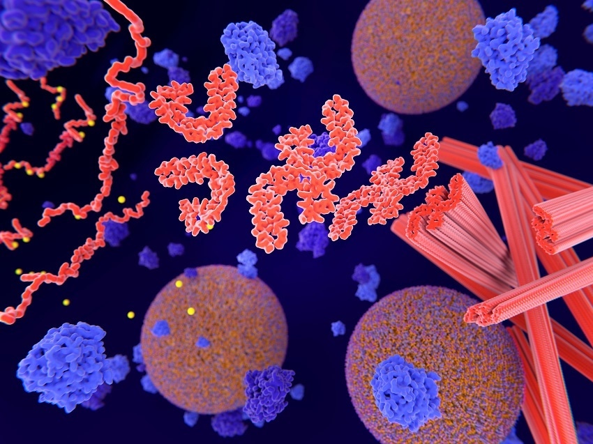
Image Credit:Shutterstock/JuanGaertner
What makes a biomolecular system complex?
A lot of these systems are based on multivalent carrier systems, which are based on macromolecules coupled with different types of peptides, antibodies, nucleic acids, drugs, and so on.
This coupling can be covalent, but mostly it is not and the non-covalent binding is the main challenge for characterization using separation methods and different detection techniques. We are working with biopolymer systems or biomacromolecular systems in which a very pronounced polydispersity exists. This means that we have big macromolecules combined with small drugs, different bioconjugates or biohybrids in different sizes, or more than two components within the one system.
These systems are, in most cases, responsive against external conditions. We have to understand how the conformation depends on the conditions and the state they attain. We also typically need to quantify the amount of drug, for example, and to understand the self-assembly processes of the systems.
What is the best method for performing conformation analysis on these complex systems?
Asymmetrical flow field flow fractionation (AF4) is an ideal method to help us to understand the properties of biomacromolecular systems. Separation using this method works according to diffusion coefficients of different sized particles or macromolecules, and low shear forces allow a broad molar mass range to be analyzed.
We can very easily change the eluent (pH) and, using AF4 in conjunction with different detectors, characterize molar mass and size distributions, as well as quantitatively characterize, for example, an amount of encapsulated drug. When AF4 is coupled with MALS and DLS detection we can obtain conformation parameters in terms of the scaling exponent as well as the shape parameter, rho.
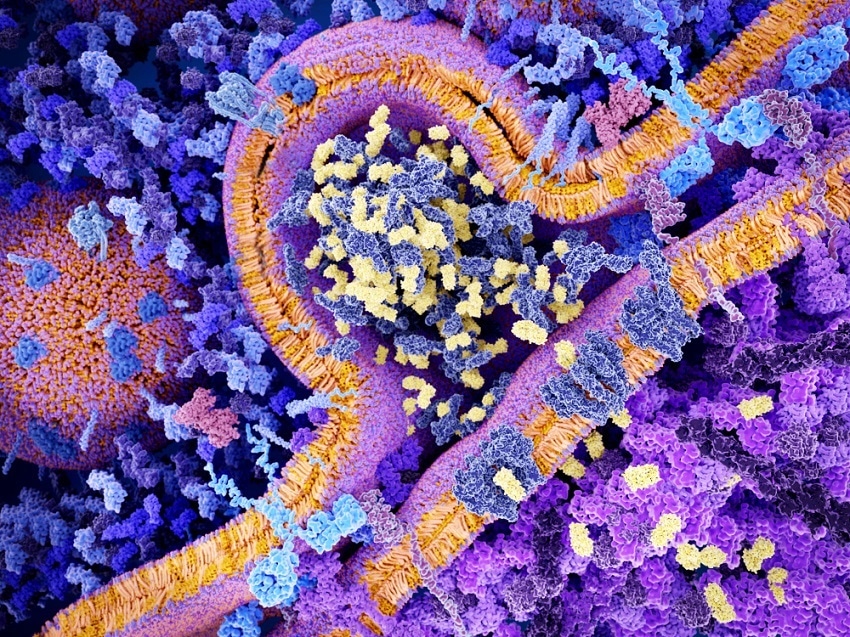
Image Credit:Shutterstock/JuanGaetner
How do you carry out quantification analysis of complex biomacromolecular systems?
We perform quantification analysis on these systems by MALS coupled with RI and UV detection.
A good example of this is the quantification of polymersomes: vesicles based on copolymeric components with hydrophobic and hydrophilic parts, similar to liposomes, which can self-assemble and encapsulate other molecules. These polymers are cross-linked so that, depending on the pH value, they can shrink or open their membrane, allowing small guest molecules to penetrate. I will use the example of myoglobin encapsulated in a polymersome which, depending on the pH value, is able to transform guaiacol into biphenoquinone.
The question appears, how many myoglobin molecules can be encapsulated within one polymersome? In order to answer this question we use AF4 coupled to MALS and UV detection in order to detect both the myoglobin and the polymersome.
Myoglobin and polymersome have very different sizes. While myoglobin is about 17,000 Daltons, polymersomes have sizes in excess of 100 nanometers. UV detection of the myoglobin shows a clear signal while the polymersome produces the same MALS signal with or without myoglobin. Therefore, by detecting the residual myoglobin which is not encapsulated into the polymersomes, we can back-calculate the amount of encapsulated polymersome.
What other applications of SEC-MALS-DLS-IV and FFF-MALS-DLS do you envision?
We use these techniques extensively to characterize dendritic and pseudo-dendritic polymers, macromolecular aggregation and self-assembly, the formation of biohybrids that couple proteins and biopolymers, and nano-encapsulation of small molecule drugs (as well as proteins). Besides these, the methods are widely applicable to many macromolecular and supromacromolecular systems, especially in the context of novel materials and nanotherapeutics.
Where can our readers go to find out more?
To find out more please visit www.wyatt.com/Polymers and www.wyatt.com/Nanoparticles.
About Albena Lederer
.jpg)
Albena Lederer is a scientist in the Analytical Department of the Institute of Macromolecular Chemistry at IPF Dresden and head of the Polymer Separation Group.
She is an extraordinary professor at the Department Chemistry and Polymer Science of the Stellenbosch University.
 Exosomes: Are They All Alike and Why Does it Matter?