|
|
|
| |

|
|
| |
The latest fluorescence news from NewsMedical |
|
|
|
| |
 How to choose the right fluorescence microscope How to choose the right fluorescence microscope
Selecting the right fluorescence microscope can be challenging, with options ranging from entry level widefield systems to advanced confocal and super resolution platforms. This Learning Centre article outlines the key technical, practical and application driven factors to consider, helping researchers make an informed choice for their imaging needs.
| |
|
|
|
|
 | | | 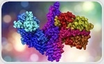 Optical methods in proteomics offer insights into protein structure and dynamics, leveraging light interactions for enhanced analysis beyond mass spectrometry. Optical methods in proteomics offer insights into protein structure and dynamics, leveraging light interactions for enhanced analysis beyond mass spectrometry. | | | | | 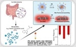 Researchers from the National Institute of Education, Nanyang Technological University, Singapore (NIE NTU, Singapore) and Singapore-MIT Alliance for Research and Technology (SMART) - Massachusetts Institute of Technology’s (MIT) research enterprise in Singapore - in collaboration with clinicians from the National University Hospital (NUH) and Yong Loo Lin School of Medicine, National University of Singapore (NUS Medicine), have developed a novel... Researchers from the National Institute of Education, Nanyang Technological University, Singapore (NIE NTU, Singapore) and Singapore-MIT Alliance for Research and Technology (SMART) - Massachusetts Institute of Technology’s (MIT) research enterprise in Singapore - in collaboration with clinicians from the National University Hospital (NUH) and Yong Loo Lin School of Medicine, National University of Singapore (NUS Medicine), have developed a novel... | | | | |  This research highlights a carbon dot biosensor for ultrasensitive E. coli DNA detection, paving the way for advancements in portable diagnostic technologies. This research highlights a carbon dot biosensor for ultrasensitive E. coli DNA detection, paving the way for advancements in portable diagnostic technologies. | |
|
|
|
| | How would you rate today's newsletter?
| |
|
|
|
| |
 |
Stay updated with the latest in health and medical news! Follow News‑Medical.net on Google News for real‑time updates. Click here to follow us now. |
| |
|
|
|
|
|