Shutterstock | Digital Photo
Raman spectroscopy is an information-rich, label-free, non-invasive imaging technique perfect for life sciences research. It uses laser light scattering in order to provide a chemical fingerprint at each point of the analyzed area and detects the molecules existing in samples.
Label-Free, Information-Rich Imaging
- Differentiate cell types and anatomical tissue layers
- Image the distribution of biomolecules in whole organisms
- Rapidly image biological samples
- Explore the chemical processes within a sample
- Minimal sample preparation
- Study chemical processes and the effects of therapeutic drugs and agents on cells
- Image biomolecule interactions taking place during cell division and other dynamic cellular processes
- Non-destructive and non-contact analysis. Does not need chemical labeling or stains
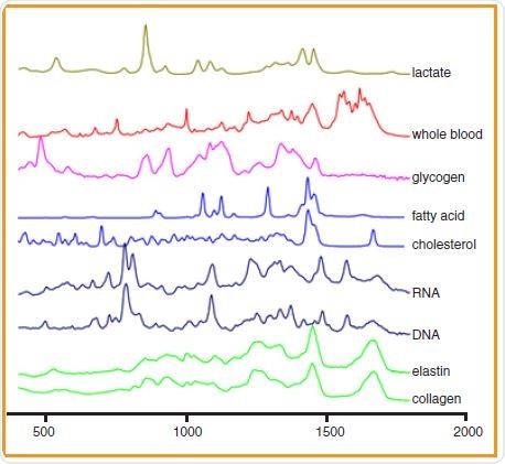
These Raman spectra demonstrate the range of different biological components that can be discriminated using Raman spectroscopy.
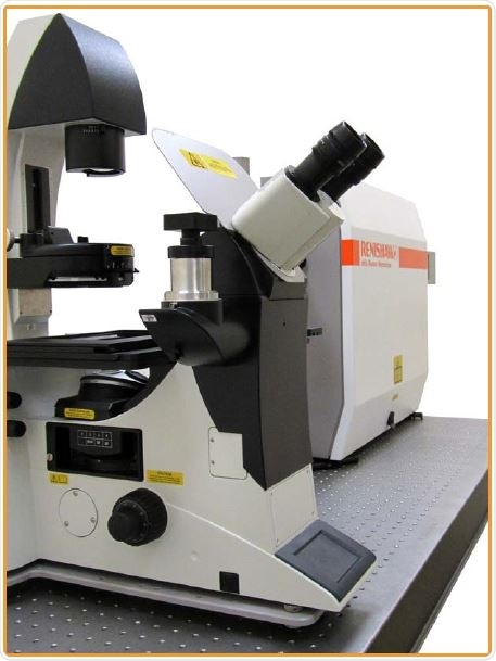
Renishaw’s inVia Raman Microscope is Ideal for Life Sciences Research
- Highly sensitive confocal Raman microscope
- Supports a variety of advanced Raman image generation techniques
- Easy-to-use live cell incubator to maintain optimal growth conditions during live cell Raman analysis
- LiveTrack for the rapid analysis of uneven biopsied samples
- Produce high spatial resolution confocal 2D and 3D images, which reveal cellular and tissue architecture. Scrutinize small features.
- Collect chemical information from variety of living tissue levels, from cell sub-organelles to whole tissue sections
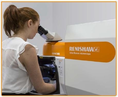
The inVia Qontor confocal Raman microscope.
Medical research using Raman Spectroscopy
Applying Innovation to Biological Imaging
Example Applications
Cell Imaging
Extract chemical information without manipulating genes, or using antibodies or stains. This helps guarantee that the results reflect the true chemistry of the cells.
- Analyze individual cells within a population and define cell-to-cell variability. For instance, examine the distribution of lipids and DNA in abnormal and healthy cells.
- Study live cells by employing a cell incubator in order to control temperature, humidity and CO2 concentration and in order to keep cells in their normal physiological states during analysis.
- Identify and then differentiate cancer cells from normal cells, stem cells from differentiated cells, and different sub-states in a cell population (example, stem and progenitor cells).
- Detect cells without known markers, based on their inherent chemical profiles. There is no need to conjugate with antibodies or manipulate genes.
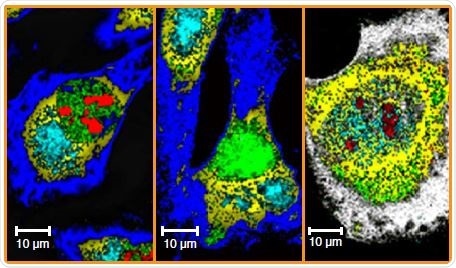
Raman image of human osteosarcoma (bone cancer) cells. Compare the size and distribution of organelles and biomolecules in normal and abnormal cells. Normal, autophagic and apoptotic MG-63 cells are shown from left to right.
Tissue Imaging
Extract a complete spectrum of chemical information (from entities such as nucleic acids, lipids and proteins) without the need for targeting biomolecules, markers, dyes or stains. Clearly visualize tissue morphology with Raman images.
- Produce Raman images whilst preventing photothermal damage to the tissue
- Perfectly distinguish, identify and then demarcate cancerous, pre-cancerous and healthy tissues, without the need for colorimetric or fluorescent labeling.
- Analyze the development of an organism, tissue’s response to stimulants or drugs, and the pathogenesis of diseases
- Study the distribution, conformation, concentration, redox and spin states, and orientation of biomolecules.
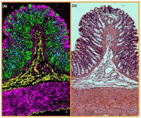
Comparison of a Raman image and hematoxylin and Eosin (H&E) stain of a healthy rat colon crypt. The Raman false-colored composite score image shows the different cell types and anatomical layers (a) differentiating connective tissue (yellow), muscle (purple), mucin (green) and nuclei (blue). Data courtesy of Riana Gaifulina, University College London, UK.
Redox Biology
Elucidate redox states within biological systems in order to study redox dynamics and its effects on diseases and health regulation. Produce Raman images in order to visualize the distribution, spatial relationships, redox and spin states of haem proteins within biological systems.
- Detect haem proteins by their Raman spectra and define their redox and spin states by Raman band positions
- Elucidate haem protein information within biological systems in situ. Raman imaging offers information on the relationship between their protein, organelle, distribution and cell functions
- Shed light on the organelle, protein and cell functions and dynamics via redox biology
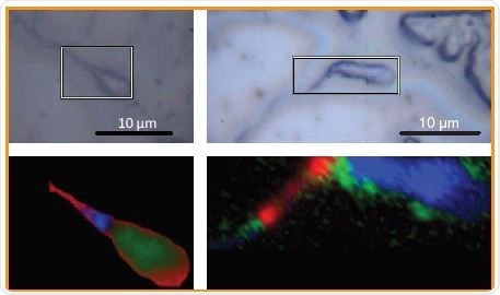
White light and Raman images of spermatozoa. In the Raman image, reduced cytochrome signals are detected in blue and red domains.
Combine Raman Spectroscopy with the Existing Biological Imaging Techniques
To obtain maximum efficiency, it is possible to analyze the sample with two or more techniques, including inverted or upright configurations, without the need for transferring the sample between instruments. With Renishaw’s correlative microscopy systems, users can be confident that they are analyzing the same point with both techniques.
Confocal Laser Scanning Microscopy (CLSM)
- Add Raman imaging to the CLSM and correlate confocal fluorescence images with Raman chemical images
- Renishaw has incorporated Raman spectroscopy with CLSM systems such as the Olympus Fluoview, the Zeiss LSM, the Leica SP8 CLSM and the Nikon A1 CLSM
Epifluorescence, Differential Interference Contrast (DIC) and Dark-field Microscopy
- Renishaw’s inVia Raman systems use research grade microscopes and are well-matched with DIC, epifluorescence and dark-field contrast imaging
- Carry out co-located imaging using fluorescence, contrast, and Raman spectroscopy
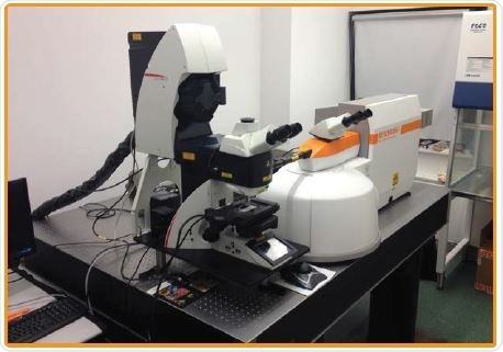
Combined Raman and confocal laser scanning microscope.
Atomic Force Microscopy (AFM)
It is possible to couple inVia’s chemical imaging capabilities to AFMs on free standing AFMs, and upright and inverted microscopes.
Standard integration packages include:
Bruker (Dimension Icon FastScan Bio and BioScope Resolve), Nanonics Imaging Ltd (MV2000 and MV4000), JPK (NanoWizard) and NT-MDT (Integra).
Inverted Microscopes
Incorporate research grade inverted microscopes and Raman spectroscopy.
The Leica DMi8 bespoke integration offers access to all ports for various other techniques. Renishaw has also developed custom solutions for Nikon, Zeiss, and Olympus inverted microscopes.
Live Cell Incubator
- The compact stage-top Okolab incubator is compatible with both upright and inverted microscope
- Control CO2, temperature and humidity
Bigger environmental chambers are also available.
No matter how unique the requirements are, Renishaw’s Special Products Team is capable of developing a custom system to meet all requirements.
inVia equipped with both inverted and upright microscopes.

This information has been sourced, reviewed and adapted from materials provided by Renishaw plc.
For more information on this source, please visit Renishaw plc.