Osteoarthritis is the leading cause of chronic disability in the elderly. In 2003, the World Health Organization revealed that it affects around 9.6% of men and 18% of women over the age of 60i. Patient treatment could be improved considerably if it was possible to detect the initial signs of cartilage degeneration earlier.
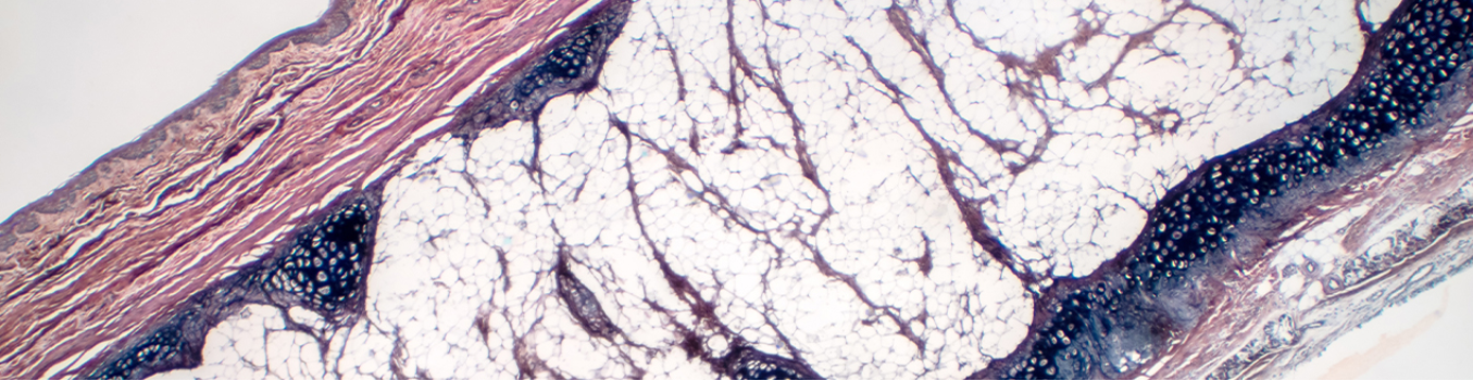
Image Credit: Renishaw plc - Spectroscopy
Recently, a team of researchers and clinicians at University College London, Royal Veterinary College and Charing Cross Hospital, led by Dr Riana Gaifulina, have launched investigations in this area. They have shown that Raman spectroscopy could help determine the grade of cartilage erosion and overall osteoarthritis disease state. This could help bring about advances that lead to earlier diagnosis and treatment of osteoarthritic degeneration.
Rapid non-invasive Raman spectroscopy can identify small molecular changes in cartilage constituents. They searched for the target molecule, sulphated glycosaminoglycan (sGAG), in cartilage samples, with a Renishaw inVia™ confocal Raman microscope to obtain single spectra across the surface of the cartilage.
This gave a detailed representative analysis of the samples, so they could evaluate the progress of cartilage degradation. The researchers could then determine the sGAG S-O stretching bond as a key element of late stage cartilage damage with the application of Renishaw's WiRE™ curve fitting algorithm to analyze the spectral range, 980 cm-1 to 1120 cm-1 and, specifically, the 1063:1003 cm-1 peak ratio.
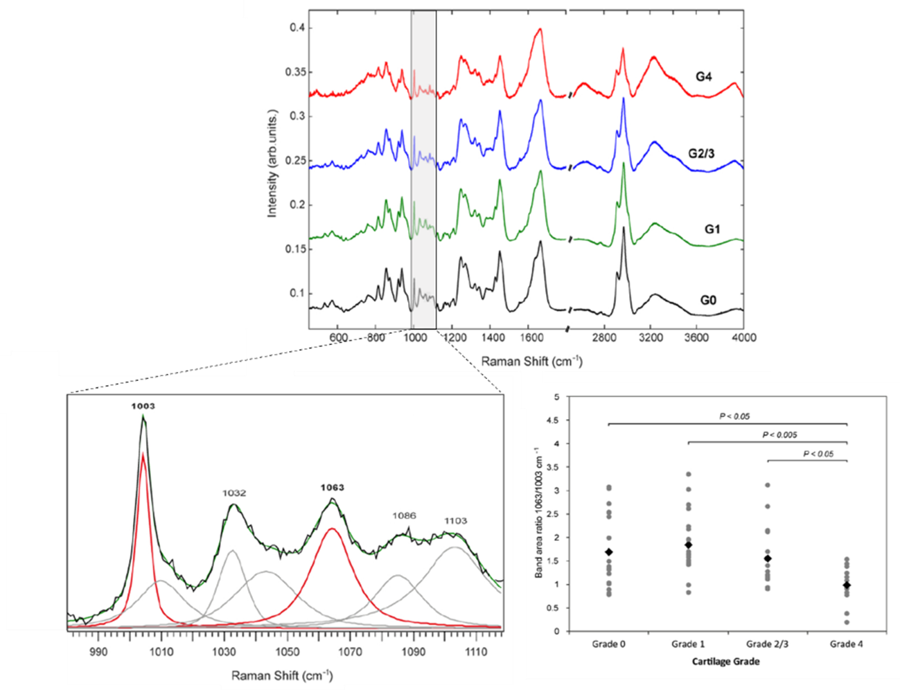
Raman spectra from Grade 0 to 4 cartilage samples. Curve fitting is employed over the 980 cm-1 to 1120 cm-1 spectral range. The 1063:1003 cm-1 peak ratio indicates a considerable difference for Grade 4 disease progression. Image Credit: Renishaw plc - Spectroscopy
The researchers also conducted principal component analysis (PCA) on both the high wavenumber and fingerprint spectral regions, and discovered that the high wavenumber region could help discriminate early disease much more effectively.
With the understanding of which peaks are indicative of disease, the researchers used the Renishaw RA816 Biological Analyzer, as this is well-suited to routine use. They quickly Raman imaged healthy and diseased areas of cartilage across multiple samples with Renishaw's fast StreamLine™ mapping technology. They discovered that Raman mapping could show the considerable differences between healthy cartilage and supposedly normal looking cartilage from a lesioned joint, in addition to the spatial distribution of cartilage constituents.
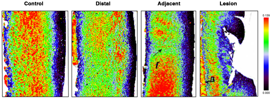
Raman maps were constructed of sGAG distribution in a healthy cartilage sample (control), and three regions (normal distal to lesion, normal adjacent to lesion, and lesion) within a lesioned cartilage sample. The diseased regions displayed a lower sGAG concentration with more heterogenous distribution. Image Credit: Renishaw plc - Spectroscopy
They also paired this with a custom-made optical probe to the inVia system. This facilitated this routine for the first time, to evaluate the potential for in vivo in-clinic Raman analysis during knee joint arthroscopy.
They were able to obtain single point spectra from visually normal and lesioned cartilage within the same joint. Interpretable spectra could be obtained in around 2 minutes. This work clearly indicates that Raman data could be captured in vivo from patients receiving knee joint arthroscopy. However, it also demonstrated that analysis of single peak ratios is generally insufficient for predictive classification and more detailed multivariate spectral analysis is necessary.
In this study the researchers demonstrated that, owing to its capability to identify small molecular changes in cartilage constituents, Raman spectroscopy could be an effective method to evaluate cartilage degeneration. This could enable clinicians to determine molecular alterations in osteoarthritic cartilage earlier which is crucial to treat this debilitating disease effectively.
For additional in-depth analysis of the experiment, please refer to the paper in the Journal of Clinical Spectroscopy using the following link: https://www.sciencedirect.com/science/article/pii/S2666054721000077
This is an open access article under the CC BY license (http://creativecommons.org/licenses/by/4.0/)
Disclaimer: The Renishaw inVia Raman microscope and RA816 Biological Analyzer have been developed for Research Use Only (RUO), not for use in diagnostic procedures. i https://www.who.int/.
About Renishaw

Renishaw is a global company and a recognised leader in Raman spectroscopy. Offering high performance optical spectroscopy products for over 20 years, Renishaw’s Spectroscopy Division is passionate about manufacturing the very best Raman products. It has a team of scientists and engineers specialising in the development, application and production of high performance, configurable Raman spectrometers.
With a foundation built on sound engineering, these systems offer the highest levels of flexibility and performance and are used by academics and industrialists to tackle analytical problems across a broad range of application areas, including life science, chemicals; materials science; pharmaceuticals; semiconductors; forensics; gemmology; antiquities; and green energy, such as photovoltaics.
Renishaw’s Raman systems are easy to use and produce repeatable, reliable data, even from challenging samples. We aim to be a long-term partner, offering quality products that meet customers’ needs, both today and into the future, backed up by expert technical and commercial support.
Sponsored Content Policy: News-Medical.net publishes articles and related content that may be derived from sources where we have existing commercial relationships, provided such content adds value to the core editorial ethos of News-Medical.Net which is to educate and inform site visitors interested in medical research, science, medical devices and treatments.