The JPK NanoWizard® V from Bruker Nano Surfaces integrates high spatio-temporal resolution with a large scan area, flexible experiment design and outstanding integration with the latest optical microscope systems. The automated setup, alignment and re-adjustment of system parameters pave the way for new opportunities for long-standing and self-regulating experiment series.
Automation
Perfecting performance, increasing productivity
Automated setup, calibration and workflow open up novel opportunities for long-term, self-regulating experiment series and complicated experimental routines.
NanoWizard V Product Video
NanoWizard V Product Video from Bruker Corporation on Vimeo.
Fast scanning
400 lines/sec
Analyze dynamic biological processes in real-time through adaptive, intelligence-based scanning routines, molecular recognition and fast force mapping.
NanoMechanics
Quantitative imaging
Nanomechanical characterization of single molecules, tissues, cells and extremely delicate samples as a result of advanced force control.
Discover the 5th generation BioAFM
NanoWizard V is anticipated to considerably progress the understanding of molecular mechanisms and dynamic cellular processes. Its PeakForce-QI mode allows quick and flexible quantitative nanomechanical measurements.
This considerably expands the abilities of AFM — while the automated, remote-control and fast-scanning capabilities offer high-throughput, high-performance imaging of even the most complicated experiments.
NanoWizard V has novel scanner and sensor technologies and advanced control software that comprises an intuitive, workflow-based graphical user interface (GUI) to guarantee true, user-friendly AFM operation.
- Unmatched user-friendliness
- Speed is available for dynamics and enhanced throughput
- Automated and high pixel density for mapping and imaging
- Proven by 8500+ publications with biological importance
- Identified by an install base of more than1000 JPK/Bruker BioAFMs globally
- From the pioneers of BioAFM with more than 25 years of experience in designing BioAFMs
- Assisted by dedicated cantilever growth for high-resolution imaging and tailored applications
- NanoWizard V leads the way for new scientific discoveries with:
- PeakForce-QI, PeakForce Tapping®, PeakForce QNM®, QI
- Single Cell Force Spectroscopy (SCFS)
- Single Molecule Force Spectroscopy (SMFS)
- New software environment V8
- DirectOverlay 2 available for AFM in conjunction with the latest optical microscopy
- Traits the new ExperimentPlanner and ExperimentControl
- Accessories available for high AFM and NA optics, environment control, and more
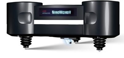
Image Credit: Bruker Nano Surfaces
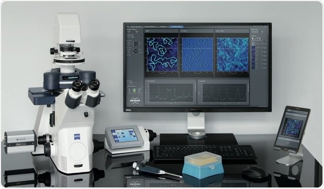
NanoWizard V BioScience setup on Zeiss Axio Observer with tablet control. Image Credit: Bruker Nano Surfaces
Latest technology for maximum efficiency
Highest performance BioAFM
The NanoWizard V features an improved and workflow-based design with innate user guidance in the new SPM V8 software. It enables beginners and experts alike to obtain higher quality reproducible data. The high degree of automation in the system enhances productivity and maximizes throughput. The latest analysis and batch processing routines guarantee scientific precision and statistical data reliability.
- Adaptive intelligent scanning routines allow quicker scanning rates of up to 400 lines/second
- Lowest noise scanner and detection system guarantee high-resolution data and unrivaled performance on inverted optical microscopes
- PeakForce-QI, the symbiosis of PeakForce Tapping and QI modes, provides the quickest, and most advanced force control for very delicate samples
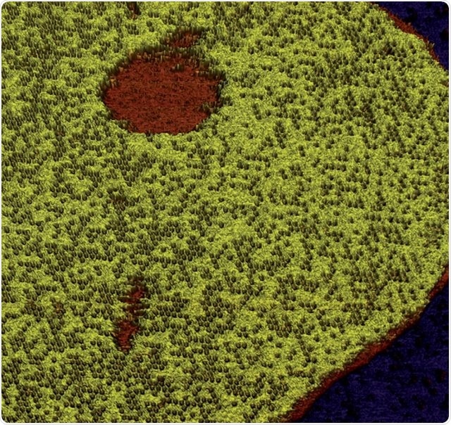
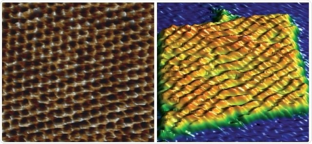
Tapping mode topography image of Annexin 5 protein on lipid membrane in buffer acquired at 40 Hz line rate showing the occupation density of centric trimers in 2D honeycomb structure. Scan size 1.3 µm × 1.3 µm, height range 8.0 nm. Image Credit: Bruker Nano Surfaces.
Fastest automated BioAFM for corrugated samples on an inverted microscope
- Users can visualize dynamics with Bruker’s Nested Scanner and new feedback technologies
- Bruker’s proven DirectDrive raises cantilever excitation stability
- Enhanced productivity and maximized throughput facilitates better statistics
- Smart optimization routines provide quantitative nanomechanics
- Active balancing offers quicker scanning over large scan ranges
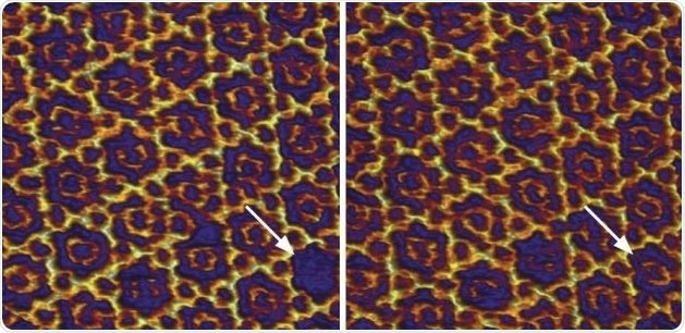
Fast tapping mode topography image acquired at 170 Hz line rate of Annexin 5 protein on lipid membrane in buffer. The arrows depict the occupation of a centric trimer from solution in the 2D crystal structure 12 seconds (8 frames) later. Scan size 96 nm × 96 nm, height range 1.0 nm. Image Credit: Bruker Nano Surfaces.
Most intuitive BioAFM operations
- Basic user-friendliness features
- User management, perfect for multi-user facilities
- Availability of automated setup and workflow
- Long-term, and unattended experimental procedures
- Remote operation abilities
- Single-click optical image calibration can be performed
- Improved storage of parameters and favorites
- Comes with intuitive integration of data processing routines
- Single-click cantilever calibration possible
- Prolonged optical viewing field available for AFM with stitching feature
Left: Topography image of atomic lattice of mica in liquid. Image taken in closed-loop on an inverted microscope, scan size 10 nm × 10 nm, height range 210 pm. Right: DNA origami (GATTA-AFM, Gattaquant, Germany) Tapping Mode topography image acquired at 400 lines/sec in TAE buffer, scan size 96 nm × 96 nm, height range 3.1 nm. Image Credit: Bruker Nano Surfaces.
The NanoWizard V is excellent for medical component analysis.”
Prof. Dr. rer. nat. Hans Bäumler, Head of Research Department, Institute of Transfusion Medicine, Charité University Hospital Berlin