An interview with Dr Bernard Siow conducted by April Cashin-Garbutt, MA (Cantab)
Can you please give an introduction to your research?
The kind of research I'm interested in is a technique called diffusion MRI and this technique is sensitive to the microstructure of the tissue. Particular clinical applications that it is used for are cancer and looking at neurodegenerative diseases.
Benefits of Compact MRI
Benefits of Compact MRI from AZoNetwork on Vimeo.
In a normal MRI scan, you can get anatomical information with a resolution of approximately 1 mm with a clinical MRI scanner, and 100 µm with a pre-clinical scanner. What diffusion MRI aims to do is look at the microstructure of the tissue with spatial information on the 1-10 µm scale.
What we're looking at, in diffusion MRI, is spatial information that is 1,000 times finer compared with clinical MRI and it's a really interesting technique to be able to elucidate what's actually going on in the tissue in terms of the microstructure.
The idea is to have some sort of non-invasive histology, enabling us to take samples without needing to stick a needle in somebody or a sample. It's an interesting technique and there's a hugely active research field surrounding it.
My particular interest associated with diffusion MRI and microstructure is looking at the permeability of the cell membrane, which is abnormal in a number of illnesses and diseases. It's also very interesting in terms of the basic physiology of the cells and basic science.
What particular techniques are you using?
We're using fairly advanced diffusion MRI techniques, coupled with computational models of tissue, which is quite a nice marrying between computer science and also MR physics. This allows us to use the data from the MRI and using the model parameters of the computational model, we can get information such as axon diameters, cell size, and cell density. This is a real up-and-coming research technique, and hopefully this will be able to move into the clinic fairly soon.
Another technique that I've been interested in is something called double-diffusion encoding. This has a wide range of potential applications. It's only recently beginning to become more popular.
One of the areas that I'm particularly interested in is looking at the permeability of cell membranes using this technique. This can be done by measuring diffusion at one time and then measuring it again. Any molecules that moved in between the two compartments would have different diffusivity.
By using this advanced technique, we are able to work out the permeability of the cell’s membrane. At the moment, this technique takes quite a long time. My efforts are trying to reduce this time into something that is clinically viable or something that can be used in a study.
What are the potential applications for these techniques?
The potential applications are in multiple sclerosis, cancer, the different inflammatory diseases and neurodegenerative diseases. As you can see, there is a very widespread potential set of applications for this technique.
It has the potential to be extremely useful for research in multiple sclerosis, because here we're looking at the permeability of the axons and in multiple sclerosis the axons become demyelinated, and as they come become more demyelinated the permeability across the myelin layers increases.
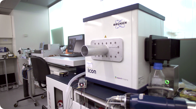
What benefits come from using the ICON?
The ICON is useful because currently we have it placed along with our nuclear imaging techniques such as our PET scanners and SPECT scanner. That has made it much easier to work within that sort of environment.
The beauty of the ICON is that it can be set up in any lab. For a traditional superconducting magnet, you need a large amount of space. A technical room with chilled water and air conditioning is required and in addition to this, a room for the magnet itself. You also need a control room.
Recently I've installed a new superconducting magnet. The cost of the building [work] for it was quite high. To be able to just wheel in a MRI scanner, like you can with the ICON, and use it, is a huge cost saving in addition to the lower capital costs of the system itself.
Since the ICON is a compact MRI system, it can be placed in any lab. It doesn't require the detailed site planning that is involved in setting up a superconducting system. For the ICON we just moved it into the lab and it just sat there quite happily. This is a huge benefit.
Being MRI experts with decades of MRI experience, how satisfied are you with the performance of the ICON?
When I first received the ICON, I was very dubious about its abilities because the signal to noise ratio, in theory, goes up with field strength and the ICON is a 1 T magnet. However, I've always worked on a 9.4 T magnet, so I thought there would be about 10 times less signal to noise, but we've not experienced this signal to noise loss. To our surprise the signal to noise ratio on the ICON was much higher than the field strength of 1 T seems to suggest, which probably is due to the efficient solenoid coil designs.
Although yes, the gold standard is the 9.4 T, and if you have the time and the budget to get a superconducting magnet, then I would go for that. However, there are some advantages to a 1 T over a 9.4 T. One of these is that the 1 T closer to clinical field strengths. This is a real benefit for some studies and we’ve had a couple of researchers that say, "Oh no, we don't want to work on the 9.4 T, we want the 1 T because it's closer to clinical field strengths."
I think the application of the ICON 1 T for pre-clinical MRI will put these scanners into many different types of labs. It's a great opportunity to grow the MRI community.
What are you doing with the ICON that you used to do with the high-field system and are the results comparable?
A great example of using the 1 T is when it outperformed the 9.4 T in a study looking at brain tumors and their localization. The contrast of the brain tumors at 1 T was really quite marked and it's actually quite difficult to achieve the same sort of contrast with 9.4 T.
In the end, what we had to do to get a similar sort of contrast in the 9.4 T, is insert an exogenous contrast agent, which is obviously less than ideal than just scanning the animals without that contrast agent, which was the case when using the ICON.
We have also done a recent study looking at liver metastasis and although we found that it's not quite as good as the 9.4 T, it certainly does the job that is required of it.
How quickly are the non-MRI experts who are using your ICON able to obtain the results that they require?
It's very simple to use, with about half an hours training and a good protocol, a non-MRI expert would be able to get the ICON up and running and would be able to be left to their own devices. This is not so true with the 9.4 T, there's many more safety aspects in regards to its use.
The fringe field of the 1 T is mostly contained within the magnet, whereas for the 9.4 T, the fringe field is much larger and it's contained within a room, and before going into that room you'd have to complete a stringent safety survey.
How has the ICON been able to help in freeing up the high-field MRI?
Three years ago our 9.4 T was never available, nearly 24 hours a day, seven days a week, it was being used. There were many studies that we couldn't do because it was so busy.
This is what lead us to the ICON, we wanted to find alternatives, and the ICON seemed like something that would be suitable. You don't need a 9.4 T scanner for every single study, sometimes it's a bit overkill. The idea was to get a low field scanner that can be used for studies that don't require such a high field scanner.
The ICON has freed up a lot of time on the 9.4 T MRI, and that has allowed more technically demanding studies to be done on the 9.4 T and now many of the other studies are now being performed on the 1 T.
How long have you had the ICON for? How long did it take to install and get it up and running?
We've had the ICON for about three years. I really don't even remember the set-up procedure - it was so quick. It was just a few days wheeling it in and testing it, and that was it. It was a very simple process. As I've said before, I've been involved in setting up a superconducting MRI scanner recently, and it was a very involved process that took months to plan.
In addition, Bruker has decades of expertise in setting up superconducting magnets and MRI scanners including their ICON and they are very helpful and very knowledgeable about the machines.
How do you plan to use the ICON in future studies?
To continue to use it for looking at brain tumors, as the contrast is so good, they're beating 9.4 T in terms of contrast. That should be a fairly large and interesting study.
One other project that's in its infancy, is my area of expertise, using the ICON for diffusion MRI. What we'll do is, again, compare and contrast the 9.4 T and 1 T for diffusion MRI studies.
 Preventing Wine Adulteration