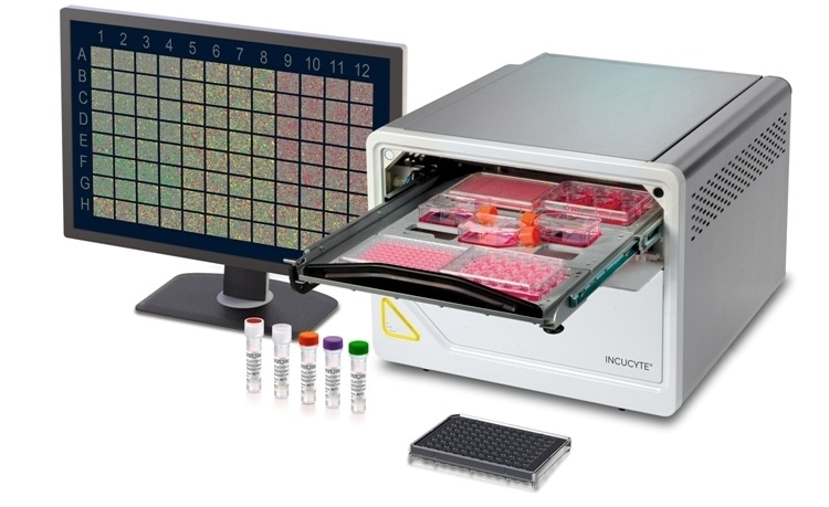
- New, patent pending optical module incorporates long wavelength NIR fluorescence to reduce phototoxicity in long term live-cell experiments
- Up to five different fluorescence channels allow researchers to explore new applications, such as metabolism, with addition of new live-cell ATP and MMP assays
- More plex, up to three fluorescence channels and HD phase in a single experiment, enables scientists to extract more data and derive more insight

Sartorius, a leading international supplier for the biopharmaceutical industry, today launched the new Incucyte SX5 Live-Cell Analysis System, the latest in the company’s line of live-cell analysis products. The Incucyte SX5 offers new capabilities, including novel optics, a new line of paired reagents, and purpose-built software to enable researchers to draw greater insight from every live cell experiment.
The Incucyte SX5 contains optics that are designed specifically for long term live-cell experiments and with flexibility in mind. Whether researchers are studying oncology, immuno-oncology, immunology or neuroscience cell models, users will be able to transition seamlessly from one question to the next. We are particularly excited to introduce the patent pending optics and analysis including NIR channel in our system, which, in contrast to the blue color channels most other systems use, significantly minimizes phototoxicity in cells.”
Kim Wicklund, PhD, Head of Product Management for the Incucyte, Sartorius
With its new optical design, the Incucyte SX5 offers up to five different fluorescence channels, up to three at a time, extending the applications and cell models that can be explored. With the unique fluorescence configuration, plus HD phase optics, more data is generated from every imaging session, enabling unique insights from complex cell models. Researchers can spend weeks preparing a cell culture or other advanced cell model for a single experiment, where different attributes like cell health, proliferation, metabolism, morphology and movement are each visualized with a different channel. Three color channels allow users to not only save time but ask more of their cell models. For instance, co-cultures are easily explored, with two channels used to identify attributes of two cell types, plus a third channel to detect health or function. And because the Incucyte SX5 can address up to six independent microplate experiments in parallel, it streamlines live-cell imaging and analysis in lab settings where multiple individuals are using the instrument concurrently.
Very sensitive cell types can be imaged and analyzed in the Incucyte SX5 for days or months without ever being removed from the incubator. The instrument keeps cell stationary and in a physiologically relevant for the duration of an experiment. All the while, integrated, user-friendly software analyzes the results in real time.
At Sartorius, our goal is to advance the biomedical field by helping every researcher conduct their dream experiments – the ones that get to the heart of a scientific question - without compromising on experimental setup or conditions. The Incucyte product line has already been featured in over 3000 peer reviewed publications. We look forward to seeing what new science is enabled by the Incucyte SX5 as our users explore the new possibilities it offers.”
Kim Wicklund, PhD, Head of Product Management for the Incucyte, Sartorius
The Incucyte SX5 will be featured today, May 5th at a free LabTools webinar, “Live-Cell Analysis in 2020: Advancing Technology to meet Researcher’s Needs,” hosted by The Scientist. Speaking at the webinar, Kim Wicklund and Dan Appledorn, PhD, Head of Product Development for Cell Imaging at Sartorius, will discuss the Incucyte and the future of live-cell analysis, and Professor Ted Hinchcliffe will discuss putting live-cell analysis into action in an oncology setting at the Hormel Institute. All are encouraged to attend.