In this interview, News Medical talks to Ben Capozzoli about the best way to prepare nuclei samples before single cell sequencing workflows and optimize nuclei extraction.
What tissues can nuclei be extracted from?
Nuclei extractions accommodate a wide variety of tissue types and preservation methods. We have had researchers use mammalian tissues from mice, rats, humans, and others preserved through snap freezing, OCT, and FFPE, as well as extracting directly from fresh tissue.
One of the main benefits of extracting nuclei for sequencing is that the method is more accommodating of different preservation methods that would make extracting viable cells impossible, like freezing the tissues or preserving them in OCT or FFPE. Therefore, banked samples from years of collection can be analyzed simultaneously through single nuclei sequencing.
Similarly, single cell suspensions that are relatively easy to generate from mammalian tissues are simply not achievable in plant tissues due to the presence of the cell wall. For this reason, researchers who wish to sequence plant tissues at a single-cell level can only resort to nuclei isolations to get a result.
How can customers optimize nuclei extraction?
Nuclei can be extracted in several ways. For example, we have used the Miltenyi extraction buffer and gentleMACS™ system, which automates the grinding process. It’s a great system for processing many samples simultaneously and ensuring that all samples are processed the same way.
Another kit we have used is the 10x Genomics kit, which involves manual grinding but is still an excellent extraction system. We have also successfully used user-formulated buffer options for a variety of different tissue types and species.
One of the most important considerations in the nuclei workflow is keeping everything cold. The cell membrane generally protects the nuclei, but they become more fragile when isolated. This means we must move quickly and keep everything cold to slow or prevent osmotic lysis of the nuclei, as we want as many nuclei to make it through to the sequencing step as possible.
My advice is to prepare everything in advance. Dilutions should be done ahead of time. Buffers should be chilled or warmed to their appropriate temperatures to ensure there is no wait time, as that will affect extraction efficiency. That information can be found in the kit's instructions.
Lastly, there will need to be filtration steps to remove as much cellular debris as possible. Different tissues might require different filter sizes.

What is the best way to prepare the extracted nuclei samples for quality control analysis?
The first step is equilibrating acridine orange and propidium iodide (AO/PI) fluorescent nucleic acid dyes to room temperature. The nuclei suspension should be kept on ice to ensure that it remains stable, but the AO/PI assay needs to be at room temperature to avoid signal loss.
Once the AO/PI is at room temperature for a few minutes, you can label the nuclei suspension with it. No incubation time is required for AO/PI. After mixing your suspension one-to-one with AO/PI, you can load it on the CellDrop Automated Cell Counter. The sample should be thoroughly mixed immediately prior to loading so that everything is resuspended before counting on the CellDrop.
 Image Credit: DeNovix
The combination of AO/PI and the fluorescent signal of the two stains removes a lot of subjectivity from counting and offers superior accuracy compared to the traditional method of using trypan blue.
What is the DeNovix CellDrop and what key features make it optimal for cell counting?
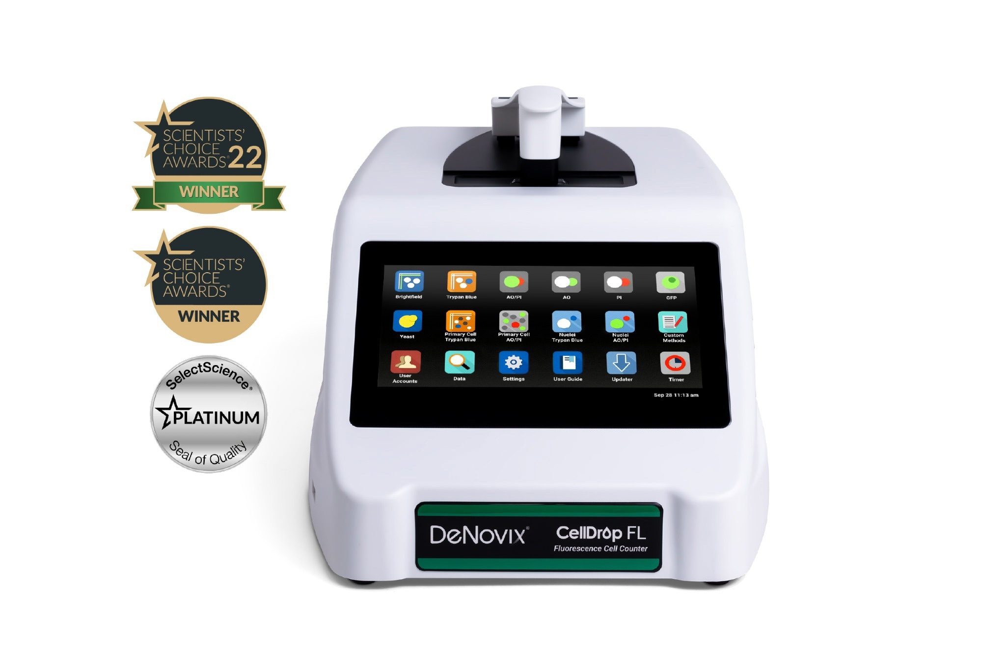
The CellDrop is a precise and reliable nuclei quantification method. It is an image-based automated cell counter with no consumables required, and it is excellent for quality control in this extraction workflow. The CellDrop takes advantage of fluorescent AO/PI labels and provides fast, reliable, and precise nuclei counts each time. The instrument has a high definition touchscreen for visualizing samples and data and can also connect to wifi and ethernet for data exporting.
The CellDrop’s patented DirectPipette™ technology requires no plastic slides, which is great for green initiatives, budget, and practicality. DirectPipette™ technology also offers performance benefits because the CellDrop uses very flat precision sapphire surfaces and a high-precision motor screw to control the arm height. Because of that, there is less variability in CellDrop measurements than you see with traditional plastic slides or hemocytometers. The sapphire surfaces also enable incredibly clear optics.
There are many features included in the software that allow the CellDrop to fit seamlessly into an established lab workflow. The counting protocol is a major tool to standardize counting across the entire lab. The CellDrop offers a variety of parameters that can be tuned from sample to sample to enable increased precision for every experimental workflow.
Once you have customized the protocol, you can use a very small volume of your overall nuclei isolation and save as much for your downstream applications as possible. These parameters are easy to set, save, and reload, which increases precision across the lab by allowing everyone to use the same count parameters.
Our applications team is happy to work with researchers to generate protocols for their specific sample type. We can provide guidance, optimize protocols, and ensure that their nuclei quality control analysis is as efficient as it can be.
What advantages does your method for imaging and counting nuclei have over traditional methods?
Traditionally, trypan blue would stain dead cells because their compromised cell membranes allow the large dye to pass into the cell. When working with nuclei, however, that cell membrane is removed during isolation. That means trypan blue will stain all the isolated nuclei dark, and the dye would be excluded by live cells. Brightfield microscopy would then be used to count nuclei.
This has been a great analysis tool for a long time and works well in clean tissue culture samples. However, trypan blue crystallizes over time. If your trypan is not clean, you could end up with debris that wastes your precious volume of nuclei and confounds your analysis.
Another issue, when dealing with blood samples, is that trypan blue cannot discern live cells from red blood cells, which we usually do not want to count. Debris will also appear dark and look like a dead cell, adding further confusion. These are all challenges presented by counting with trypan blue that render the method more subjective than fluorescence.
We recommend that our customers use AO/PI for counting nuclei. Acridine orange (AO) and propidium iodide (PI) are fluorescent nucleic acid dyes. AO is actively transported into live cells and fluoresces green. PI behaves similarly to trypan blue in that live cells exclude the dye due to its large size, but cells with a compromised cell membrane undergoing apoptosis include the dye in the nucleus and fluoresce red.
Both stains require nucleic acid binding to fluoresce. Since the nuclei isolation process removes the cell membrane, any nucleus extracted from a cell should light up red from the PI, and any cell that was not correctly lysed in our extraction will stain green. Objects that do not have a nucleus, like a red blood cell or any debris left behind from your extraction, do not fluoresce.
Fluorescence is a really powerful analysis tool that takes a lot of subjectivity out of the process by essentially turning the counting process into a binary choice of red or green.
 Image Credit: D. Kucharski K. Kucharska/Shutterstock.com
How does the CellDrop help customers achieve success in their downstream applications?
The CellDrop counting process is quick and efficient, allowing users to rapidly check the quality and quantity of a sample before moving on with downstream applications. Our customers have used this technology on different nuclei isolation protocols, using samples such as fresh kidney, frozen kidney, frozen lung, fresh tomato root, and frozen mouse brain.
One great feature of the CellDrop is that it enables you to analyze visually for debris in real-time. This will allow you to quickly determine if you need further filtration or purification before moving on with library prep. If the level of debris is too high to move on to library prep, you can use a purification kit to purify the nuclei. As CellDrop requires very little sample volume, re-checking after purification is also possible.
Once your isolated samples meet the quality control criteria, you can move on to downstream applications, such as RNA sequencing, ATAC sequencing, or flow cytometry.
These downstream applications all require a sample with plenty of nuclei, very little debris, and few intact cells. Using the CellDrop, you will know the exact concentration of your nuclei, the level of debris, and the number of intact cells in your sample. This provides confidence that you have a high-quality sample that will hopefully give you good results when you load them into your downstream applications.
Can you summarize why your customers benefit from the CellDrop?
Nuclei extractions are lengthy, sensitive, and expensive procedures. A lot of expensive equipment goes into the extractions, and that is just to isolate the nuclei, not to process those nuclei with RNA-seq or Assay for Transposase-Accessible Chromatin using sequencing (ATAC-Seq).
You need high-quality samples before you can run one of those expensive experiments, so sample quality control is a critical component of the process. Quality control will help ensure that you are not wasting money on a sample that did not extract well or had another issue that could be easily visualized by counting.
With image-based counting on the CellDrop, we can reproducibly quality control all your nuclei samples in a way that is easy to interpret, powerful, and allows you to make quick decisions before you go on with your experiment.
Where can our readers find more information?
Please visit denovix.com for additional resources. A series of eBooks and webinars cover counting methods and counting protocols on the CellDrop, and there are technical notes for different applications, including nuclei isolation applications.
I would encourage researchers to schedule a virtual demo or a free trial with someone on the application science team to learn how CellDrop can improve their nuclei preparation workflow.
DeNovix also has a free trial program in the US. During the trial, researchers can work with a member of the application science team to ask any questions and tune the equipment for their specific workflow and samples. Once satisfied with their ability to count and quality control their sample on the CellDrop, researchers can pack it up and mail it back. It’s a great way to test out the equipment, and it doesn’t cost anything.
About the speaker
Ben Capozzoli brings expertise in immunology, imaging, cell biology, and cell counting to the DeNovix team. After receiving his BS in Biology from Susquehanna University, Ben held a research assistant position at the University of Pennsylvania studying the effects of aging on D. melanogaster stem cells. He then went on to receive his Masters in Immunology from Villanova University, his research focusing on immuno-oncology. Ben joined the DeNovix team as an Application Scientist in 2019, and since then he has contributed to the development and application support of the CellDrop Automated Cell Counter.
About DeNovix, Inc.
WELCOME TO DENOVIX
Award-Winning products for Life Science
Our multi-award winning products include the Reviewers’ Choice Life Science Product of the Year and Platinum Seal awarded- DS-11 Series Spectrophotometer / Fluorometer and CellDrop™ Automated Cell Counter. CellDrop is the first instrument of its kind to Count Cells Without Slides. These powerful instruments integrate patented DeNovix technology with easy-to-use software designed by life scientists for life scientists.
Researchers tell us they love the industry leading performance, smart-phone-like operation, and the flexible connectivity of the instruments. When support is needed, the DeNovix team is here to help. DeNovix received the prestigious Life Sciences Customer Service of the Year based on independent reviews posted by scientists worldwide!
CellDrop: Sustainable laboratory product of the year
The CellDrop Automated Cell Counter has been awarded Sustainable Laboratory Product of the Year in the SelectScience® Scientists’ Choice Awards®!
CellDrop’s patented DirectPipette™ technology distinguishes it as the only cell counter to eliminate the need for cell counting slides. This innovation saves millions of single-use plastic slides from use and disposal each year.
Sponsored Content Policy: News-Medical.net publishes articles and related content that may be derived from sources where we have existing commercial relationships, provided such content adds value to the core editorial ethos of News-Medical.Net which is to educate and inform site visitors interested in medical research, science, medical devices and treatments.