Introduction
UV-visible range microspectrosopy is one of the most useful techniques for studying individual protein crystals. Today, protein crystals are used for bioseparations, controlled drug delivery, drug design, and structural biology. Hence, this technique is employed for studying the optical properties of individual crystals and also for performing quality control of crystals after they are grown.
Analysis of Biocrystalline Materials
CRAIC Technologies offers the 20/30 PV™ microspectrophotometer that enables easy, quick and non-destructive identification, qualification and analysis of biocrystalline materials.
The 20/30 PV™ was configured for fluorescence and transmittance for this series of experiments. The 15x Schwarzchild type objective proved suitable for this application as it has spectral consistency and ultra-long working distance over the entire visible, UV and NIR regions.
Experimental Framework
In this series of experiments, absorbance microspectroscopy was used to analyze four samples. The first sample included DNA crystals, the second had Flavoprotein crystals, the third contained salt crystals in solution, and the fourth included DNA-Protein crystals.
From each sample, 200 µl of solution was removed and placed in a Costar 96 well plate since the well plate exhibits excellent UV transmission characteristics. Then, a crystal was identified and Kohler illumination was used to optimize the system.
Before acquiring the crystal spectrum, a reference was run off to the side of each crystal. This process removes the spectral characteristics of the solution, the well plate, and the instrument. The final spectrum is of the crystal itself.
Measurement of Absorbance Spectra
Following this, the absorbance spectra of the four samples were measured, as shown in Figure 1.
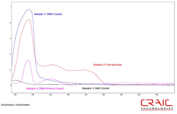
Figure 1. Overlay of absorbance spectra of Samples 1 to 4.
All the four samples were labeled as a DNA crystal, flavoprotein, salt crystal in solution, and DNA/protein crystal, respectively. With the exception of salt, the spectra in all the crystals showed an absorbance peak at 286nm. In the DNA crystal, this was the only peak observed and was extremely intense specifying a high optical density.
Upon initial examination, shoulders were also exhibited by the flavoprotein at 354, 414 442 and 446 nm. However, on repeated examination, a change was observed in the crystals spectrum, as shown in Figure 2. The DNA/Protein crystal i.e. the fourth sample also showed a peak at 352nm.
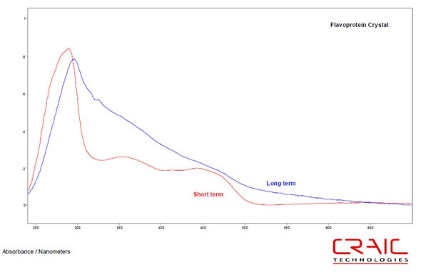
Figure 2. Overlay of flavoprotein crystal absorbance spectra at short and longer term exposure to oxygen.
Images were also obtained for each crystal. Figure 3 shows the four samples, the DNA crystal, the flavoprotein crystal after it starts dissolving, the salt crystal in solution, and the protein/DNA crystal. The black square is the entrance aperture of the microspectrophotometer; the aperture was 15x15µm in these images.
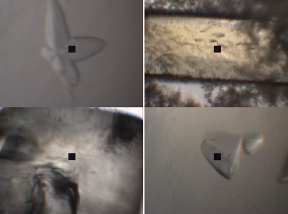
Figure 3. Images of the four samples
Conclusion
In these experiments, UV-visible absorbance spectra were acquired from a DNA crystal, DNA/protein crystal, flavoprotein crystal, and salt crystal. The spectra in all the crystals can easily be identified and differentiated from one another. The optical densities, and thus the concentrations can also be compared. Moreover, the spectral change in the flavoprotein can also be monitored.
Acknowledgement
Produced from articles authored by Dr. Paul Martin, CRAIC Technologies, Inc.
About CRAIC Technologies
 CRAIC Technologies™ specializes in developing superior UV-visible-NIR microanalysis solutions: we build instrument to collect spectra and images of sample features ranging from sub-micron to hundreds of microns. CRAIC Technologies products include UV and NIR microscopes, UV-visible-NIR microspectrophotometers, instruments to measure thin film thickness and colorimetry on the microscopic scale, Raman microspectrometers, automation solutions, traceable standards and more.
Sponsored Content Policy: News-Medical.net publishes articles and related content that may be derived from sources where we have existing commercial relationships, provided such content adds value to the core editorial ethos of News-Medical.Net which is to educate and inform site visitors interested in medical research, science, medical devices and treatments.