Sponsored Content by PittconNov 24 2015
The need for accurate cancer detection
The ability to distinguish cancer cells from healthy cells during surgery is essential for preventing the removal of healthy tissue and ensuring no tumor remnants are left behind.
Although the need for full tumor resection is an unmet need in various cancer types, it is particularly urgent in the field of brain cancer. Suboptimal results are often seen in patients who undergo brain surgery for high-grade tumors, with tumor recurrence often occurring close to the resection cavity. Furthermore, the removal of healthy brain tissue can have extremely detrimental consequences for these patients.
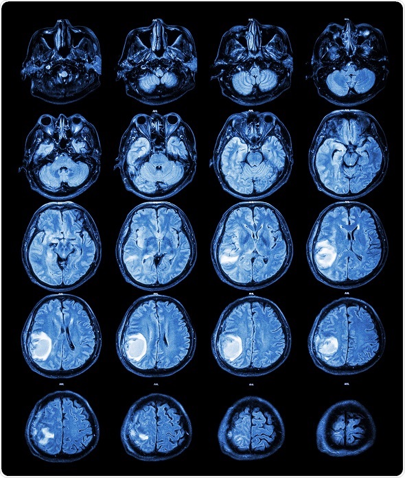
MRI: Brain tumour at right parietal lobe.
© Puwadol Jaturawuttichai / Shutterstock.com
Challenges in cancer detection
Achieving maximal tumor removal without damaging healthy tissue is a challenge in tumor surgery because surgeons cannot rely on any visual cues to indicate whether tumor resection is complete. Neurosurgeons rely on their judgment while in the operating room, but often have to make a guess as to exactly where the edges of a tumor lie.
In contrast, it is easy to differentiate between tumor tissue and non-cancerous tissue at the microscopic level. However, traditional histopathologic techniques involve tissue being removed from the operative field and sent off for processing and staining, meaning these methods cannot be performed in situ or in real-time. An in vivo technique for identifying resection margins during surgical procedures would therefore be highly desirable.
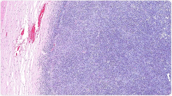
Microscopic view of oligodendroglioma of the brain (right half blue) and normal brain (pink portion on the left). The brain cancer has invaded and replaced normal healthy brain tissue. Glioma of the brain. © vetpathologist / Shutterstock.com
Stimulated Raman scattering microscopy
Now, a new laser-based microscopic technique is being developed that may help surgeons differentiate between tumor tissue and healthy tissue in real-time. Stimulated Raman scattering (SRS) microscopy is a non-invasive imaging technique that is based on the scattering of light by molecular vibrations.
Images collected using SRS microscopy show that normal brain tissue has sparse cells that contain bundles of axons, whereas cancerous tissue is full of cells organized in a disordered fashion. SRS microscopy has the potential to be used in a wide range of biomedical applications, providing instant tissue examination, without the need for histological processing and staining. A brain area of 2x2 cm2 can be mapped in as little as 2.5 minutes, allowing for real-time, in vivo studies
In a study of the technology by Ji et al (2015), SRS microscopy was used to image brain tissue from patients undergoing surgery for glioblastoma. The technique generates signals for proteins and lipids, which are then given a color (blue and green, respectively). This allows the users to distinguish between the brain cortex, tumor tissue and white matter. SRS microscopy of biopsies taken from the patients revealed distinctive features and the presence of infiltrating cells in tissues that had appeared healthy when conventional histological staining was used.
The researchers also designed an objective classifier that combined various image characteristics such as axonal density and the protein-to-lipid ratio into one output that could alert the pathologist if tumor infiltration was occurring. The classifier could differentiate between infiltrated and non-infiltrated regions with an accuracy of more than 99%.
Neurosurgeon Daniel Orringer who co-piloted the technology says their SRS microscopy technique allows the surgical decision making process to become data driven rather than being based on a surgeon’s best guess.
While the researchers continue to test the SRS microscope, they are busy developing a second-generation instrument that can easily be operated during surgery. Surgeons could insert a fresh tissue sample into the device to produce images that would help determine on-the-spot whether further surgery is required.
In another study, Lu and colleagues (2015) performed DNA imaging using SRS microscopy to explore the technique as a non-invasive way of diagnosing skin cancer. First, they imaged a tissue sample using traditional histological staining and compared the images with those obtained using SRS microscopy.
They then imaged skin cancer tissue from patients with squamous cell carcinoma and found an increased amount of mitotic features, indicating the hallmarks of cancer – increased cell division and proliferation.
The results showed that the SRS imaging method was comparable to conventional staining techniques used in histological diagnosis. The researchers say they expect that SRS microscopy may not only speed up surgical procedures, but also have potential as an in vivo non-invasive technique for skin cancer diagnosis and the evaluation of cancer cell aggression.
Symposia at Pittcon 2016
This year’s Pittcon, the world’s largest annual conference and exposition for laboratory science, will take place between the 6th and 10th March 2016 in Atlanta, Georgia. With an impressive technical program, the event will feature more than 2000 presentations. Among these, topics will include the use of SRS microscopy as a non-invasive technique for providing instant tissue imaging in cancer patients.
We will hear about how SRS microscopy enables tumor and non-tumor tissue to be clearly distinguished and how the technology could become an invaluable tool in the operating theatre. The use of SRS microscopy to study DNA distribution and condensation in the nuclei will also be discussed, along with the technique’s potential in for evaluating skin lesions in real-time.
Among the 683 organizations exhibiting at this year’s Pittcon is Renishaw Inc., the world leader in high-sensitivity confocal Raman instrumentation, with systems based on optical microscopes and advanced fiber optic probes. Renishaw’s Raman instruments can currently be found in many cutting edge research laboratories and production environments.
Another exhibitor who will be present is B&W Tek, an advanced instrumentation company that provides Raman spectroscopy solutions through user-friendly platforms. This company has been a leading provider of Raman spectroscopy solutions for the biomedical, pharmaceutical, physical and chemical and research communities.
Also exhibiting at Pittcon is Kaiser Optical Systems, a world leading specialist in the design and production of Raman analyzers and spectrographic instrumentation.
References
- Lu F, et al. Label-free DNA imaging in vivo with stimulated Raman scattering microscopy. Proceedings of the National Academy of Sciences 2015;112:11624–11629
- Ji M, et al. Detection of human brain tumor infiltration with quantitative stimulated Raman scattering microscopy. Science Translational Medicine 2015;7:pp. 309ra163
- Notingher I, et al. Raman spectroscopy for medical diagnostics - From in-vitro biofluid assays to in-vivo cancer detection. Advanced Drug Delivery Reviews 2015;89:121–134
- Freudinger C. Fiber Scanning Stimulated Raman Scattering (SRS) Endoscope. Grantome. Available at: http://grantome.com/grant/NIH/R43-CA189320-01A1
About Pittcon
Pittcon is North America’s largest conference and exposition in laboratory science, attracting thousands of industry, academia, and government attendees and exhibitors from around the world in. Pittcon goes beyond the typical conference; it's a global community dedicated to advancing science through education, collaboration, and philanthropy. By bringing together scientists, researchers, educators, students, and industry leaders, Pittcon creates opportunities to learn, connect, and inspire innovation. Beyond the event itself, more than 90% of Pittcon's net proceeds support science education and outreach initiatives locally and globally, funding scholarships, grants, STEM programs, laboratory improvements, and other efforts that strengthen communities and expand access to science.
Pittcon delivers world-class educational and professional development opportunities through its Technical Program, Short Courses, and Networking Workshops and Roundtables. Attendees can explore the latest scientific research through symposia, oral and poster presentations, enhance their skills with expert-led deep-dive courses, and connect with peers through interactive workshops and networking experiences designed to foster collaboration and career growth.
The Pittcon Expo brings the scientific community together in an immersive environment where attendees can discover the latest laboratory technologies, meet with leading instrument and service providers, and experience science in action. Beyond the exhibit booths, the Expo Floor features engaging attractions, educational experiences, demonstrations, and networking spaces, while receptions, parties, and social events create opportunities to build lasting connections with colleagues from around the world.
Sponsored Content Policy: News-Medical.net publishes articles and related content that may be derived from sources where we have existing commercial relationships, provided such content adds value to the core editorial ethos of News-Medical.net, which is to educate and inform site visitors interested in medical research, science, medical devices and treatments.