Sponsored Content by TissueGnosticsReviewed by Maria OsipovaApr 8 2024
3D organoids serve as invaluable tools in medical research, enhancing our comprehension of intra- and inter-cellular mechanisms within tissues. Yet, their full potential in the medical field is often hindered by significant technical challenges associated with detecting and quantifying organoids.
The newly launched StrataQuest Organoid App aims to overcome these challenges by automatically identifying organoids, thus enabling the monitoring of their growth under diverse conditions. Consequently, it allows users to quantify the count, dimensions, and density of organoids within co-cultures that include immune cells.
This development grants users the capacity to conduct in-depth investigations into the intricacies of the immune system, translational medicine, oncology, precision medicine, and beyond.
From 2D to 3D cell line cultures
For many years, the bulk of human cell research has predominantly relied on 2D cell line cultures, largely because of their relative affordability and ease of use.
While 2D cell line cultures have achieved considerable success in multiple areas of medical research, including the identification of new druggable targets, their foundational creation is highly inefficient. This process requires significant genetic and phenotypic modifications to enable their survival under culture conditions.1,2
Additionally, nearly all the differentiated cell types and tissue microenvironments found in the original tissue are absent in 2D cell line cultures, meaning these models are not able to truly replicate complex in vivo processes.3 This creates a profound limitation regarding their use in tissue studies and personalized medicine.
Today, with human organoids – 3D cell line cultures constructed from stem cells that are comprised of organ-specific cell models that self-organize via cell sorting and spatially restricted lineage4 – scientists are able to replicate the architecture and physiology of human organs with astounding detail.
The ability of 3D organoid cell lines to autonomously organize and accurately resemble the cellular composition and structure of human tissue makes them a highly attractive technique for medical research.
They are extremely useful for analyzing therapeutical properties relating to immune disorders, organ development, oncology, host-pathogen interactions, wound healing, and drug development, and look to pave the way for precision therapy.5
Co-cultured organoids
Co-culturing organoids with the cells from their surrounding tissue enhances the depth of analysis for the physiological processes that occur between organoids and neighboring cell subsets, such as immune cells, stromal cells, and parenchymal cells.6
This approach offers researchers greater insight into how neighboring cells impact organoid development, leveraging both time-lapse and end-point analysis techniques by utilizing brightfield imaging.
However, determining organoid size, number, and shape is still extremely challenging because:
- Organoids enclosed in extracellular matrix gels grow at various focal depths, which makes it difficult to obtain clear focus with fixed microscopy.
- Organoids differ in size and shape due to various stages of differentiation, leading to heterogeneity.
- Dense clusters of proliferating immune cells can interrupt imaging signatures, which in turn can lead to false positives.5
Imaging-processing algorithms have been predominantly developed from the analysis of organoids without additional cell subsets, and until now, no high-throughput image analysis pipeline could determine and quantify co-cultured organoids with the appropriate accuracy. 5
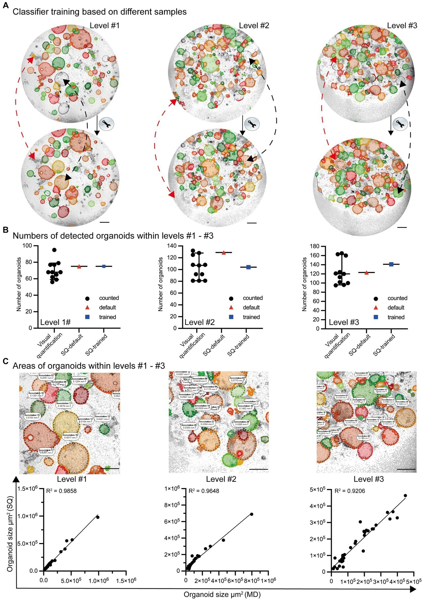
Image Credit: Stüve, P., Nerb, B., Harrer, S., Wuttke, M., Feuerer, M., Junger, H., Eggenhofer, E., Lungu, B., Laslau, S. and Ritter, U. 2023. Analysis of organoid and immune cell co-cultures by machine learning-empowered image cytometry. Frontiers in Medicine. 10, p.1274482
Examining co-cultured organoids with the organoid app
To tackle the associated issues with co-cultured organoid evaluation, a team of scientists developed the Organoid App. The App offers reliable and efficient high-throughput validation and quantification for organoids co-cultured alongside immune cells.
This groundbreaking technique will enable scientists to explore extremely complex questions with respect to organoid development and the effects immune cell subsets and other compounds have.
The StrataQuest-supported App was developed to improve the automated identification and quantification of organoid structures in lymphocyte co-cultures.
The machine learning algorithms within the Organoid App were able to identify and classify 3D organoids with co-cultures of dense immune cells with incredible accuracy, replicating that of the human eye.5
The StrataQuest software has the capacity to handle various image configurations and formats, enabling detailed analysis of co-cultured organoid development from time-lapse to end-point investigations.
The end-point analysis is performed without impedance through automation on the TissueFAXSiPLUS microscopy platform. The method involves a combination of automated imaging with sophisticated image processing techniques, including contrast enhancement, greyscale conversion, membrane detection, and structure separation. 5
Specifically, it incorporates a classifier engine designed for machine learning on the available samples.
In this approach, a grayscale image serves as the input, and users contribute data on object classes by outlining the organoid contours. The machine learning algorithm then utilizes this detailed information to generate more accurate outputs.5
StrataQuest is a comprehensive image analysis software suitable for an array of applications, encompassing both brightfield and fluorescence imaging. It offers compatibility with the diverse imaging formats of widely used imaging platforms, allowing users to export results in various file formats, including PDF, XLS, and XLSX. This flexibility facilitates both graphic and statistical analysis of the results.
The scientists also compared the Organoid App with Incucyte® software and OrganoSeg. Incucyte could not detect the differences in organoid growth between untreated and Epidermal Growth Factor-treated co-cultures, while OrganoSeg had very limited organoid detection. This comparison underscored the unique effectiveness and quality of the Organoid App’s performance for such analysis.5
Conclusion
The Organoid App, backed by StataQuest, stands out as an exceptionally useful tool for the identification and quantification of organoids in cell subset cultures, a task that was not feasible before. With its recent development, this App has the potential to revolutionize the medical research landscape, especially in the realm of translational medicine.
StrataQuest provides a variety of customized Apps designed to streamline navigation and analysis workflows, developed through close collaboration with its clients.
References and further reading
- Schutgens, F. and Clevers, H. 2020. Human organoids: tools for understanding biology and treating diseases. Annual Review of Pathology: Mechanisms of Disease. 15, pp.211-234.
- Kim, J., Koo, B.K. and Knoblich, J.A., 2020. Human organoids: model systems for human biology and medicine. Nature Reviews Molecular Cell Biology. 21(10), pp.571-584.
- Abdul, L., Xu, J., Sotra, A., Chaudary, A., Gao, J., Rajasekar, S., Anvari, N., Mahyar, H. and Zhang, B. 2022. D-CryptO: deep learning-based analysis of colon organoid morphology from brightfield images. Lab on a Chip. 22(21), pp.4118-4128.
- Lancaster, M.A. and Knoblich, J.A. 2014. Organogenesis in a dish: modeling development and disease using organoid technologies. Science. 345(6194), p.1247125.
- Stüve, P., Nerb, B., Harrer, S., Wuttke, M., Feuerer, M., Junger, H., Eggenhofer, E., Lungu, B., Laslau, S. and Ritter, U. 2023. Analysis of organoid and immune cell co-cultures by machine learning-empowered image cytometry. Frontiers in Medicine. 10, p.1274482.
- Huch, M., Gehart, H., Van Boxtel, R., Hamer, K., Blokzijl, F., Verstegen, M.M., Ellis, E., Van Wenum, M., Fuchs, S.A., de Ligt, J. and van de Wetering, M. 2015. Long-term culture of genome-stable bipotent stem cells from adult human liver. Cell. 160(1), pp.299-312.
About TissueGnostics
TissueGnostics (TG) is an Austrian company focusing on integrated solutions for high content and/or high throughput scanning and analysis of biomedical, veterinary, natural sciences, and technical microscopy samples.
TG has been founded by scientists from the Vienna University Hospital (AKH) in 2003. It is now a globally active company with subsidiaries in the EU, the USA, and China, and customers in 30 countries.
TissueGnostics portfolio
TG scanning systems are currently based on versatile automated microscopy systems with or without image analysis capabilities. We strive to provide cutting-edge technology solutions, such as multispectral imaging and context-based image analysis as well as established features like Z-Stacking and Extended Focus. This is combined with a strong emphasis on automation, ease of use of all solutions, and the production of publication-ready data.
The TG systems offer integrated workflows, i.e. scan and analysis, for digital slides or images of tissue sections, Tissue Microarrays (TMA), cell culture monolayers, smears, and other samples on slides and oversized slides, in Microtiter plates, Petri dishes and specialized sample containers. TG also provides dedicated workflows for FISH, CISH, and other dot structures.
TG analysis software apart from being integrated into full systems is fully standalone capable and supports a wide variety of scanner image formats as well as digital images taken with any microscope.
TG also provides routine hematology scanning and analysis systems for peripheral blood, bone marrow, and body fluids.
TG cooperations
TG continuously cooperates with research groups and other companies in the industry to provide novel tools and applications to its customers.
Sponsored Content Policy: News-Medical.net publishes articles and related content that may be derived from sources where we have existing commercial relationships, provided such content adds value to the core editorial ethos of News-Medical.Net which is to educate and inform site visitors interested in medical research, science, medical devices and treatments.