MAGNETOM Free.Max breaks barriers to expand the reach of MRI.
- Where access to MRI was not viable, MAGNETOM Free.Max makes access to MRI affordable
- Where siting was an obstacle, MAGNETOM Free.Max offers a helium-free infrastructure
- Where conventions have limited our thinking, MAGNETOM Free.Max breaks barriers to explore new clinical opportunities
- Where patients have felt discomfort, MAGNETOM Free.Max is the world’s first 80 cm bore offering an improved patient experience
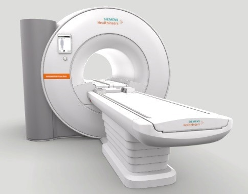
Image Credit: Siemens Healthineers
Expand opportunities with a larger bore
Bore size should not prevent patients from having an MRI. However, whether it is due to claustrophobia or a larger body, sometimes it does happen. For a more comfortable and less daunting experience, a larger bore size would help. Opening up the bore means that there is better access and new opportunities.
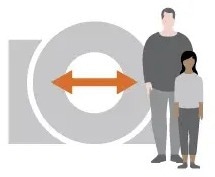
Image Credit: Siemens Healthineers
Improve siting flexibility
It is hard to install an MRI scanner in a compact space, and doing it affordably is an even bigger challenge. Traditional MRI scanners are big and bulky and often need extensive infrastructure that normally comprises a quench pipe.
A smaller, lighter MRI system with a helium-independent infrastructure would mean higher flexibility for several institutions. Minimal intensive installation needs would also simplify site selection and open up MRI to a larger pool of people.
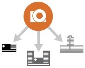
Image Credit: Siemens Healthineers
Simplify operation to make MRI easier to use
The need for exclusively trained technologists has grown as MRI exams have increased in complexity and popularity. A smarter MRI scanner would allow technologists of any capacity to achieve top-grade images via intuitive and user-friendly interfaces.

Image Credit: Siemens Healthineers
Increase the value delivered and make MRI accessible for all
Smaller, roomier, lighter, and easier to operate and install, helium-independent MRIs provide the anticipated clinical outcomes while making MR more accessible.
In the coming months, Siemens Healthineers will demonstrate how the system can also offer value for the providers, patients, and institutions it serves.

Image Credit: Siemens Healthineers
When barriers are left behind, new opportunities arise
- High-V-MRI – Value beyond barriers
- Infrastructure radically simplified
- See things like never before
- Intuitive operation for any operation
- Diagnostic confidence for daily excellence
- Get connected to stay one step ahead
- The first 80 cm patient bore
- Redefining MRI affordability
High-V MRI – Value beyond barriers
Digitalization is rapidly transforming MR imaging through the application of highly efficient acquisition techniques and deep learning-based reconstruction. High-V MRI takes the power of digitalization and deliberately applies it to a new field strength of 0.55T with inherent clinical benefits.
High-V MRI combines the best of both worlds to offer a new era in MRI that embraces diagnostic confidence in daily routine and new clinical opportunities.
The digital transformation of MRI
Siemens Healthineers is helping to kickstart the digital revolution in MRI practice.
Siemens Healthineers is continuously perfecting its acquisition technologies to attain greater image quality. Currently, with Deep Resolve, the company has introduced deep learning-based reconstruction to improve the quality of results even further.
MAGNETOM Free.Max leverages the complete potential of the digital power of Siemens Healthineers for the best diagnostic quality.
Deep Resolve Gain
Deep Resolve Gain, a constructive technique, utilizes individual quantitative noise maps in the reconstruction process for targeted denoising. It increases image quality to ensure the best diagnostic abilities and is available for a wide range of sequences.
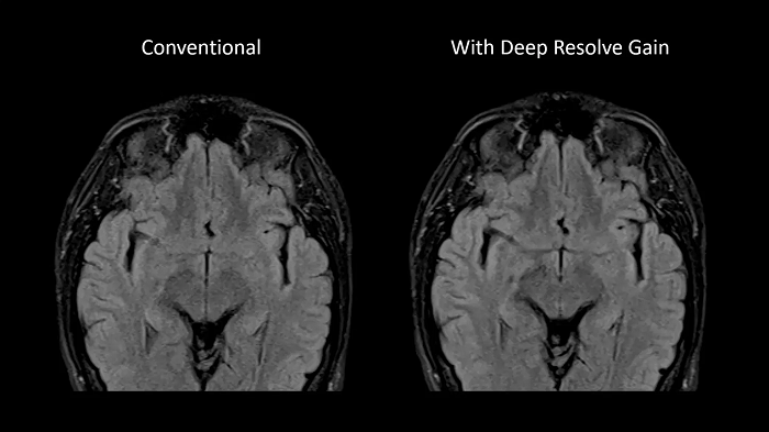
Image Courtesy: University Hospital Erlangen, Germany | Image-ID: 4aaaa0297
Deep Resolve Sharp
Using a convolutional neural network of TSE sequences, Deep Resolve Sharp produces high-resolution images from low-resolution input. Sharper and crisper images can be achieved without compromising on data acquisition time.
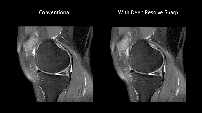
Image Courtesy: University Hospital Erlangen, Germany | Image-ID: 4aaaa0459
Simultaneous Multi-Slice
Siemen Healthineers’ Simultaneous Multi-Slice TSE is an exclusive approach to accelerate data acquisition in TSE imaging. By getting multiple slices simultaneously, users can save a maximum of 50% scan time, thereby reducing motion artifacts, minimizing patient slot times, and maintaining patients friendliness.
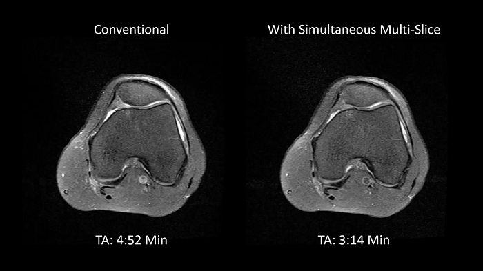
Image Courtesy: University Hospital Erlangen, Germany | Image-ID: 4aaaa0375
See things like never before
New opportunities arise when traditions are broken. The unique field strength of 0.55T with High-V MRI provides inherent physical advantages that overcome the limitations of today’s MR imaging.
MAGNETOM Free.Max offers exciting new clinical applications that break the barriers of MR physics and expand the reach of MRI.
Improved implant imaging with High-V MRI
The imaging of metal implants has historically been difficult with conventional MRI systems as metal causes artifacts. High-V MRI offers intrinsic physical advantages that result in reduced metal distortions and strongly improved diagnostic capabilities for implant imaging.
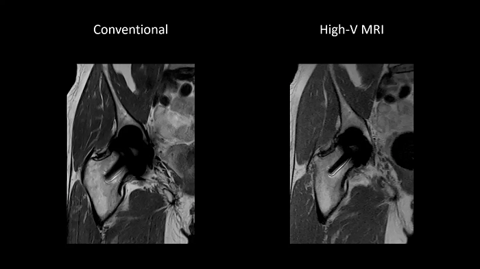
Image Courtesy: University Hospital Erlangen, Germany | Image-ID: 1aaaa3623 | 4aaaa0399
Reduced susceptibility challenges with High-V MRI
In MRI, susceptibility artifacts are a familiar phenomenon.
One notable example of such artifacts is at air-tissue interfaces, as they occur at the sinuses and orbits. The unique field strength of High-V MRI offers physical advantages that reduce susceptibility artifacts. This leads to reduced geometric distortions in diffusion imaging which results in improved diagnostic quality.
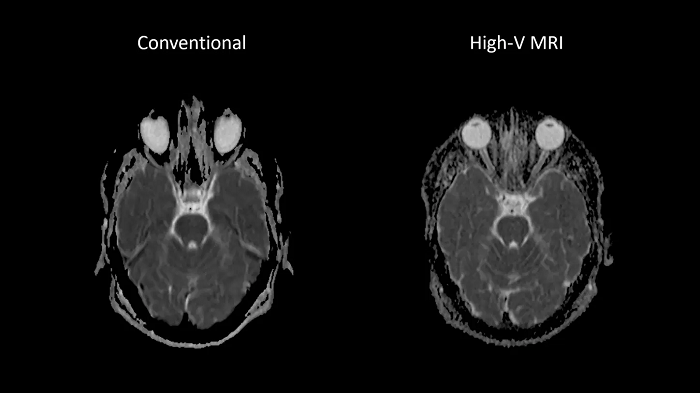
Image Courtesy: University Hospital Erlangen, Germany | Image-ID: 1aaaa3439 | 4aaaa0297
New opportunities in pulmonary imaging with High-V MRI
Pulmonary imaging has been disreputably challenging with traditional MRI as the air-tissue interfaces lead to fast signal decay. These challenges scale with magnetic field strength which makes High-V MRI the perfect opportunity for pulmonary imaging. Consequently, High-V MRI has the ability to extend the reach of pulmonary MRI.
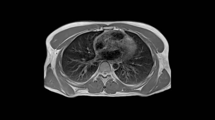
Image Courtesy: University Hospital Erlangen, Germany | Image-ID: 4aaaa0419
Diagnostic confidence for daily excellence
MAGNETOM Free.Max is first-in-class in terms of image quality. This translates into unparalleled diagnostic confidence for users’ daily routines. Driven by the company’s unique innovations in deep learning-based reconstruction and image acquisition, MAGNETOM Free.Max offers outstanding diagnostic quality for standard clinical MRI applications.
Head
EPI-based 3D SWI
A new approach towards susceptibility-weighted imaging founded on 3D EPI allows highly resolved and high-quality depiction of cranial veins on MAGNETOM Free.Max.
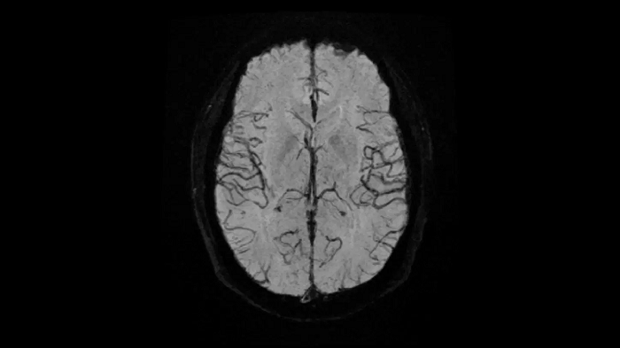
Image Courtesy: University Hospital Erlangen, Germany | Image-ID: 4aaaa0297
Time-of-flight angiography
Non-contrast enhanced angiography of the cranial vessels is used to detect vessel occlusions or aneurysms. High-resolution ToF angiography can also show small arteries for subtle diagnoses.
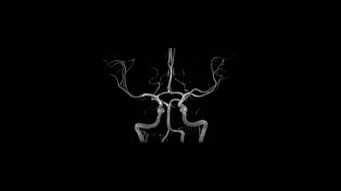
Image Courtesy: University Hospital Erlangen, Germany | Image-ID: 4aaaa0466
Neck
Excellent soft tissue contrast in the neck
All-inclusive soft tissue examination of the head with outstanding image quality for T1, T2, and diffusion contrast.
- Head/Neck Coil
- Spine Coil
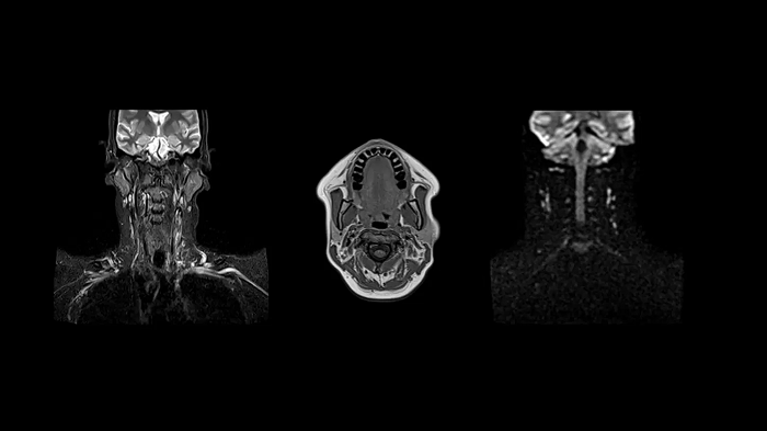
Image Courtesy: University Hospital Erlangen, Germany | Image-ID: 4aaaa0458
Spine
C-Spine - T2 TSE
Integrate Head/Neck and Spine Coil to gain outstanding C-Spine images with T2 TSE.
- Head/Neck Coil
- Spine Coil
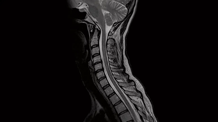
Image Courtesy: University Hospital Erlangen, Germany | Image-ID: 4aaaa0440
L-Spine - T1 TSE
Optimal depiction of the lower spine with T1 TSE imaging using the Spine Coil.
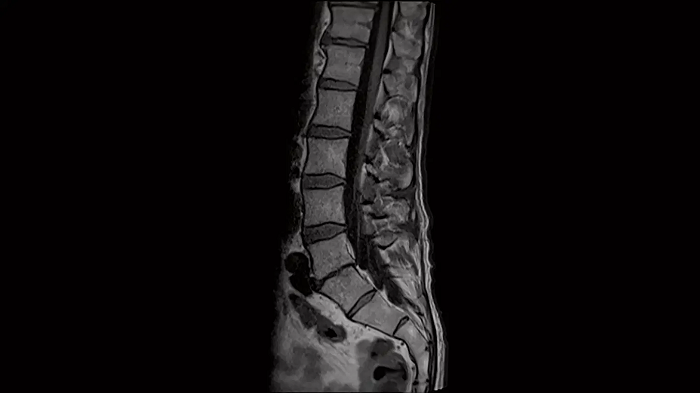
Image Courtesy: University Hospital Erlangen, Germany | Image-ID: 4aaaa0376
Shoulder
Shoulder - PD TSE FatSat
Exceptional fat suppression for the clear imaging of the shoulder.
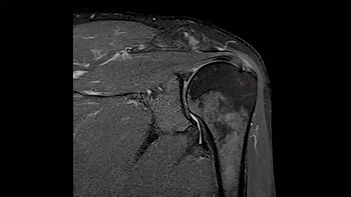
Image Courtesy: University Hospital Erlangen, Germany | Image-ID: 4aaaa0384
Abdomen
MRCP with Compressed Sensing
Abdominal imaging with T2w SPACE of the biliary ducts can be expedited with Compressed Sensing to reduce acquisition times. Both techniques offer high-resolution, high-quality MRCP for uncompromised abdominal investigation.
Compressed Sensing factor 6.5 | 1.2 x 1.2 x 1.0 mm3 | TA = 4:21 minutes
- Contour L Coil
- Spine Coil
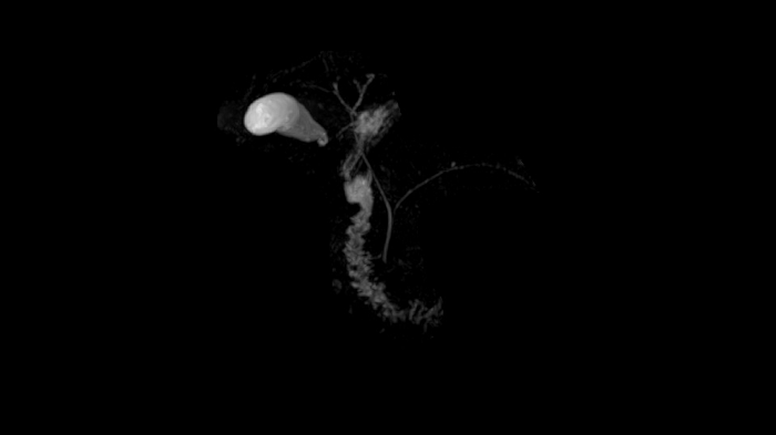
Image Courtesy: University Hospital Erlangen, Germany | Image-ID: 4aaaa0465
T1 VIBE Dixon with CAIPIRINHA
Benefit from Siemens Healthineers’ dedicated CAIPIRINHA acceleration to gain a complete abdominal volume within a single breath-hold. The Dixon technique offers outstanding fat-water separation for a flawless representation of the abdominal anatomy.
- Contour L Coil
- Spine Coil
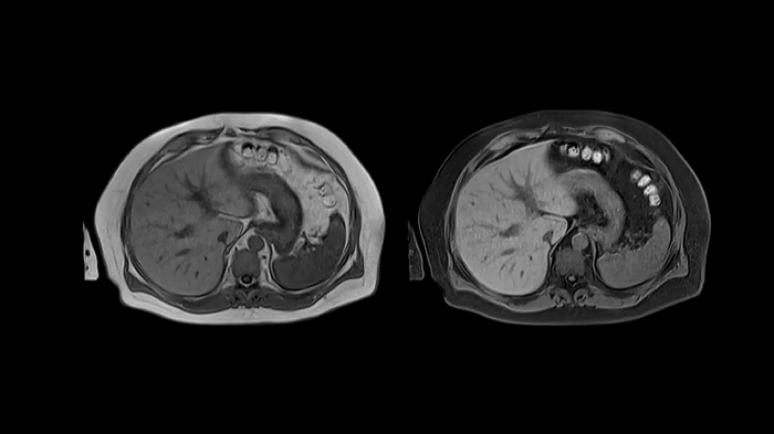
Image Courtesy: University Hospital Erlangen, Germany | Image-ID: 4aaaa0458
Body diffusion imaging
Outstanding quality diffusion imaging for comprehensive abdominal MR capabilities.
- Contour L Coil
- Spine Coil
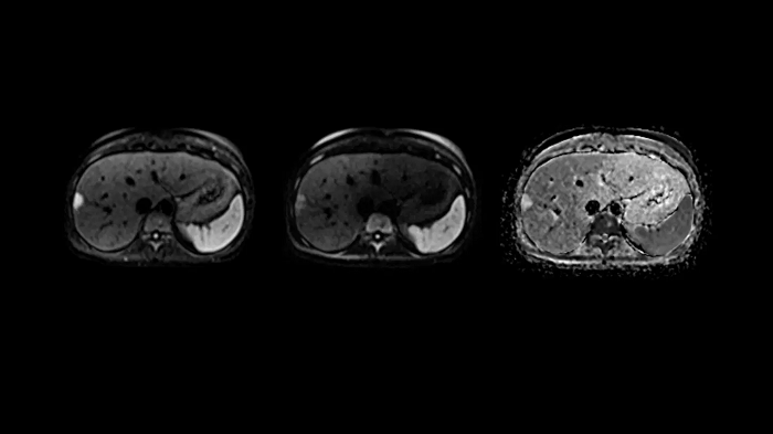
Image Courtesy: University Hospital Erlangen, Germany | Image-ID: 4aaaa0339
T2 BLADE
T2 motion insensitive abdominal imaging using BLADE results in outstanding outcomes, regardless of the orientation or if fat saturation is needed.
- Contour L Coil
- Spine Coil
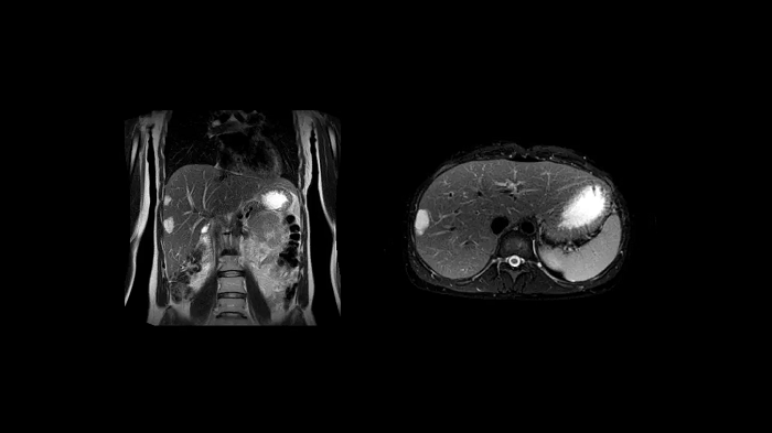
Image Courtesy: University Hospital Erlangen, Germany | Image-ID: 4aaaa0339
Knee
PD TSE FatSat with SMS
In MSK imaging, TSE is the workhorse. Using the Siemens Healthineers dedicated SMS TSE technique, users can stay on top of measurement times without compromising on image quality while resolving orthopedic clinical concerns.
TA = 3:46 minutes
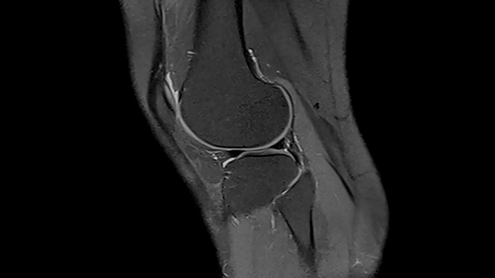
Image Courtesy: University Hospital Erlangen, Germany | Image-ID: 4aaaa0449
Ankle
Ankle - PD TSE FatSat
Even in demanding anatomies such as ankle with Contour S Coil, achieve optimal image quality.
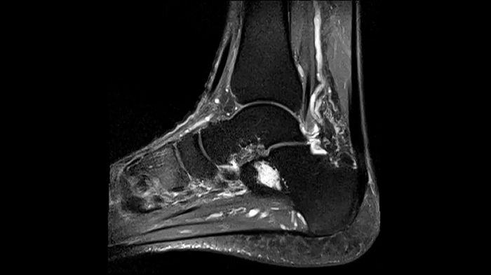
Image Courtesy: University Hospital Erlangen, Germany | Image-ID: 4aaaa0381
The first 80 cm patient bore
The first step of patient care is putting the patient at ease. Using the first and only 80 cm patient bore, MAGNETOM Free.Max overcomes limitations associated with accessibility and patient comfort, thereby making the experience a realistic choice for anxious, claustrophobic, and more corpulent patients.
The first 80 cm wide bore redefines patient accessibility
The 80 cm wide bore of MAGNETOM Free.Max in comparison to conventional bore sizes. Image Credit: Siemens Healthineers
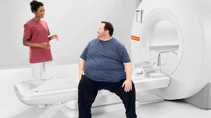
The extra wide bore provides greatly improved access for bariatric patients. Image Credit: Siemens Healthineers
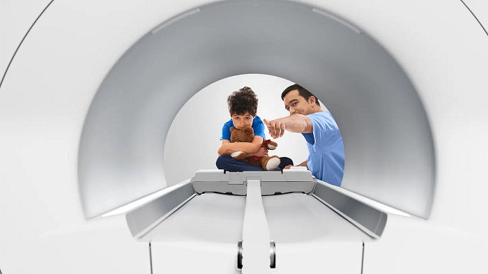
The 80 cm patient bore strongly reduces anxiety and increases patient comfort. Image Credit: Siemens Healthineers
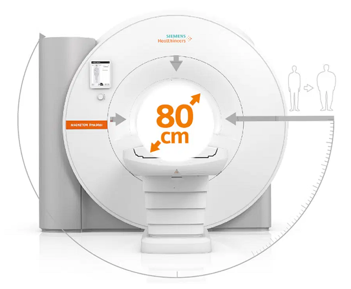
Extra wide bore with 80 cm. Image Credit: Siemens Healthineers
Infrastructure radically simplified
Only the need for MRI should define where it is located. MAGNETOM Free.Max is Siemens most compact whole-body MRI, and with DryCool technology, it provides a helium-independent infrastructure.
MAGNETOM Free.Max overwhelmingly simplifies infrastructure requirements, breaking down the walls imposed by siting constraints.
Intuitive operation for any professional
Using myExam Companion, MAGNETOM Free.Max eliminated the barriers associated with MRI operations. Thanks to the new possibilities of AI and digitalization, data is turned into integrated expertise and tailored assistance to benefit the user and address the clinical question.
Enter the era of intuitive MRI with BioMatrix Technology and syngo Virtual Cockpit.
Explore the streamlined MRI workflow with MAGNETOM Free.Max
myExam Companion
MRI is intelligently automated by myExam Autopilot. Even amateur users can produce high-quality outcomes seamlessly in routine MRI scans. This also allows experienced technicians to concentrate more on patients instead of scanner operations. myExam Cockpit and myExamAssist complement the scan experience using flexible protocol customization and guidance.
BioMatrix
MAGENTOM.Free.Max simplifies users’ tasks beyond scanning. Users can achieve reproducibility and efficiency when setting up a patient using BioMatrix workflow automation.
syngo Virtual Cockpit
MAGENTOM.Free.Max directly integrates syngo Virtual Cockpit remote assistance into users’ workplaces. Benefit from expert advice whenever it is needed.
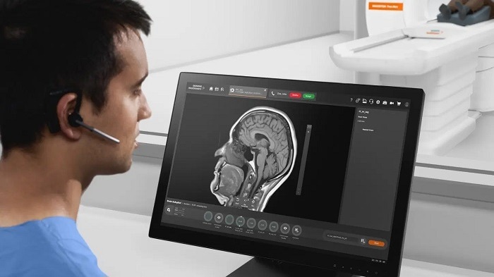
Image Credit: Siemens Healthineers
Redefining MRI affordability
A viable business seeks to expand the reach of its MRI. MAGNETOM Free.Max breaks down financial barriers, opening up new opportunities when it comes to offering MRI at the front line of diagnostic services.
Stay connected to be one step ahead
Users can focus on caring for their patients, as Siemens stay connected to care for their MRI. Siemens Healthineers’ innovative service uses the preventive intelligence of the Guardian Program to solve technical issues before downtimes occur. Siemens stays connected to keep users one step ahead.