ParaVision 360 is one of the most sophisticated preclinical imaging software currently available.
Highlights
- Consistent quantification: The highest accuracy in all research stages, from set-up to quantification, from day to day, and from subject to subject, leading to the most consistent, reliable data.
- Greatest imaging power: An extensive and inventive selection of methodologies, paired with advanced tools for viewing, reconstructing, and analyzing, delivers unparalleled insights from imaging subjects.
- Seamless multimodal imaging: Consistency in MRI, PET, and µCT for user-friendly application.
Focusing on consistent quantification, ParaVision 360 makes scanning even more intuitive while maintaining the full flexibility that Bruker users expect. Bruker's exclusive, expanded MRI sequence portfolio, along with its robust image data evaluation capabilities, leads to maximum proficiency in scanning and analysis. At the same time, the simplicity and uniformity across the range of instrumentation allow operators to focus on their research.
ParaVision 360 V3.5 is now available
Highlights from the newest features of the 3.5 release:
Out-of-the-box protocols for morphological, metabolic, and molecular studies
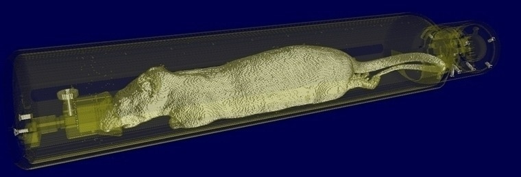
- Fully automated PET/CT stitched protocol, including attenuation correction (AC) map for rats up to 250 mm in length
- Standardized rat T2* EPI protocol for fMRI meeting guidelines of the MultiRat collaboration
- Fast, T2-weighted 3D, isotropic in vivo imaging with RAREvfl
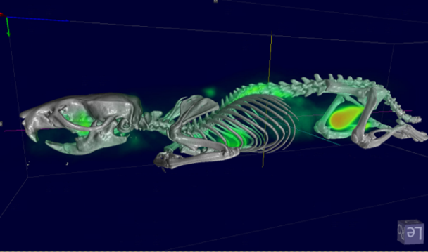
Fast extended PET/CT imaging for full rat body coverage. Image Credit: Bruker BioSpin Group
Extended spectroscopy capabilites
- AI-based baseline and phase correction
- Efficient visualization and processing of single-voxel spectroscopy and chemical shift imaging spectroscopic data (e.g., CSI) with Spectroscopy Card
- Metabolic map creation and anatomical reference image overlaying
- Spectral editing of J-coupled resonances such as lactate, γ-aminobutyric acid (GABA), glutamate, and glx (glutamate/glutamine) with MEGA-PRESS
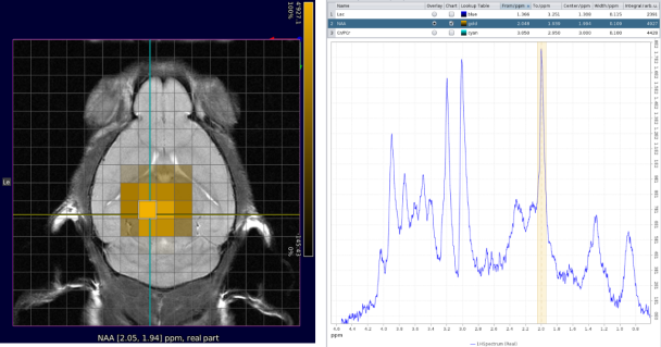
CSI with NAA frequency mapped. Image Credit: Bruker BioSpin Group
Efficient PET scanning
- Data is displayed on the touchscreen as well as in pre-defined single slices for fastest planning of further scans based on tracer biodistribution
- Real-time 3D display of tracer biodistribution during scanning
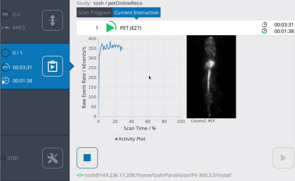
Touchscreen display of count rate evolution and MIP online reconstruction. Image Credit: Bruker BioSpin Group
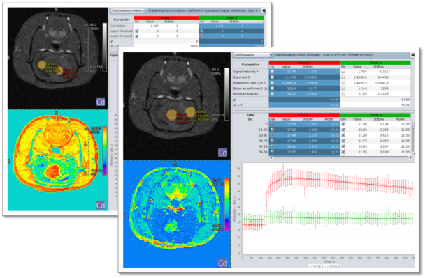
Interactive navigation, including immediate update of parameter map. Image Credit: Bruker BioSpin Group
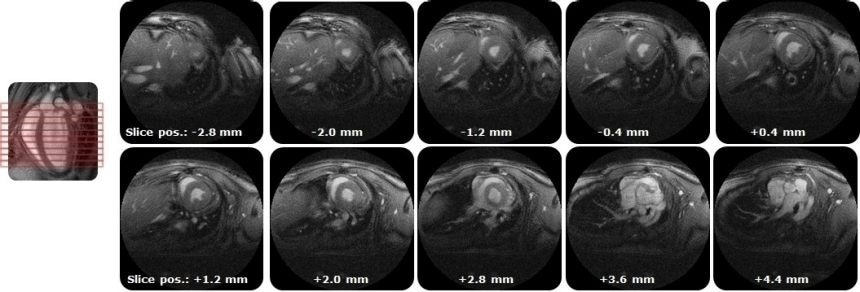
IntraGateUTE, with its radial readout, enables high-quality cardiac cine data in shortest measurement time. Image Credit: Bruker BioSpin Group
Automatic MR-based Attenuation Correction (AC), calculating for animal cradle, MRI RF coil, and full animal body. Image Credit: Bruker BioSpin Group
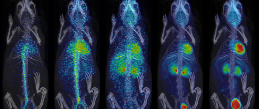
Dynamic 18FDG/PET scan in healthy mouse acquired in LM, reconstructed with variable intervals and displaying myocardium and blood. Image Credit: Bruker BioSpin Group
Cross-modality highlights
- Advanced intensity based image registration
- Animal Transport System control via touchscreen1
- Automatic 3D image fusion
- An Acceleration Suite including Compressed Sensing, Simultaneous Multislice Imaging, Partial Fourier with homodyne reconstruction, and 3D GRAPPA with CAIPIRINHA
- Common terminology between instruments, common integrated multimodal workflows, and common image processing, viewing, and analysis tools and functionalities for optimal clarity
- Data browser with simple search functions for accessing stored protocols and recorded data
- Database and archiving and report creation
- Drag and drop image processing
- Multi-station image acquisition and imaging stitching1
- MR Method development framework
- Magnet status display in ParaVision and remote instrument monitoring option
- On-the-fly quantitative image mapping
- ROIs with thresholding, seeded region growing, and freehand definition
- Simple selection of prone or supine viewing
- SLASER single voxel spectroscopy that is robust against B1 inhomogeneities and provides less chemical shift displacement
- Wobble control on magnet front touchscreen display²
1Available for BioSpec 3T, Inline PET/MR, and PET/CT Si78
2For instruments with Animal Transport System
Highlights for MRI
- 3D GRAPPA and Compressed Sensing2
- B1 optimization and mapping
- Dynamic shimming capability1
- Fat-water separation imaging
- Fat chemical shift corrected images
- Fast online decisions and real-time calculations on all channels
- IntraGateUTE for virtually flow-artifact-free cardiac scanning
- MRI pulse program display
- MRI CASL workflow package for optimal scanning and consistency
- Over 100 validated and ready-to-use in vivo protocols and scan programs for mice and rats with integrated examination guide for optimal scanning navigation and reproducibility
1 Work in progress research feature. Outcomes depend on the available shim hardware
2 Compressed Sensing is work in progress
Highlights for PET
- Advanced DOI
- Custom isotope profiles
- Extensive reconstruction algorithms (e.g., MLEM, OSEM, MAP, FBP)
- Kinetic time course image reconstruction and segmentation
- MRI or CT-based attenuation corrections with automatic loading of animal cradle and MRI coil and individual calculation of subject
- Multimodal Scan Programs include complete instructions for interdependent PET/CT reconstruction steps
- TAC display with Patlak fit for kinetic modeling
- Scatter, randoms, isotope decay, and dead-time corrections
- Static, multi-stage, dynamic imaging
- Wireless cardiac PET/MR imaging
Highlights for µCT
- 3D/2D
- Cardiac and respiratory gating
- Hounsfield scaling
- Protocol library for mice and rats for anatomical reference
- Static, multi-stage, dynamic imaging
- Step & shoot or continuous
- Smoothing, beam hardening & ring corrections
- Real-time adjustments preview
- Ultrafast low-dose protocols