axoCells™ Human iPSC-Derived Microglia, male donor, ≥1 million cells
Microglia, the primary immune cells in the brain, play vital roles in neurogenesis, homeostasis maintenance, synaptic plasticity, and brain development. They are widely used in monoculture for compound screening and co-culture with neurons and muscle cells to model AD and ALS.
- Derived from human iPSCs
- Assay ready in just 7 days
- Express the key markers including IBA-1, TMEM119 and P2RY12
- Demonstrate robust functional activity, measured by cytokine release, phagocytosis and chemotaxis
- Used in advanced in vitro models including co-culture and tri-culture
Get the Full Guide on Human iPSC-derived MICROGLIA
Phenotypic characterization
Axol Bioscience has meticulously characterized its axoCells Microglia, confirming precise morphology and expression of crucial cell markers through immunocytochemistry and flow cytometry. To maintain high quality and consistency at scale and support reliable in vitro models of neurodegenerative diseases, Axol manufactures them in accordance with ISO 9001 standards.
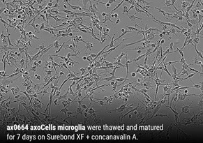
Figure 1. axoCells Microglia showing expected morphology, with different phenotypes demonstrating sub-populations with specified functions. Image Credit: Axol Bioscience Ltd
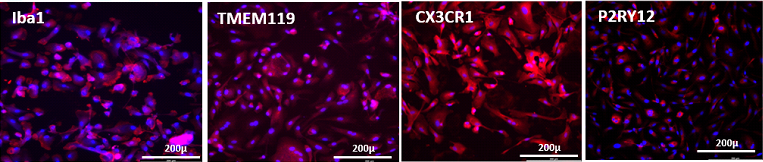
Figure 2. Immunocytochemistry of axoCells Microglia demonstrating the expression of key markers Iba1, TMEM119, CX3CR1 and P2RY12. Image Credit: Axol Bioscience Ltd
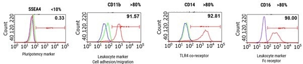
Figure 3. Example flow cytometry QC for fresh macrophage progenitors, demonstrating the presence of lineage-specific markers CD14, CD11b, and CD16 above threshold levels, and negative control SSEA4 below threshold. This batch would, therefore, pass this stage of QC. Our standard panel also includes CD206 and CD163 as lineage-specific markers. Unstained, Isotype (control), Marker of interest. Image Credit: Axol Bioscience Ltd
Get the Full Guide on Human iPSC-derived MICROGLIA
Functional characterization
To evaluate the functional performance of the axoCells Microglia, Axol conducted various assays, such as phagocytosis, chemotaxis, and cytokine release, as shown in Figure 4, 5 and 6.
Phagocytosis
axoCells Microglia can phagocytose various baits, such as beta-amyloid, S. aureus, and myelin basic protein. Cytochalasin D inhibits this activity.
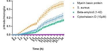
Figure 4. axoCells Microglia were thawed and matured for seven days before addition of pHrodo labelled bait. Baits were added to the cells and phagocytosis monitored over 24 hours using an IncuCyte S3, showing a steady increase over time. Cytochalasin D (10 µM) was used as a negative control and showed complete inhibition of phagocytosis. Image Credit: Axol Bioscience Ltd
Chemotaxis
axoCells Microglia respond to different concentrations of C5a with the anticipated chemotaxis, with very high concentrations having the anticipated inhibitory effect.
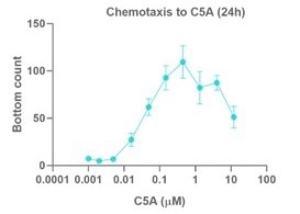
Figure 5. Chemotaxis by fresh axoCells Microglia to various concentrations of C5a. iPSC-derived microglia were matured for 7 days before re-plating into chemotaxis plates. Cell movement was measured using an IncuCyte S3. Values represent the number of cells moving from the top chamber to the bottom, towards C5a, after 24 hours. Data are n = 4 +/- SEM. Image Credit: Axol Bioscience Ltd
Cytokine release
Microglia from axoCells release the expected pattern of functional cytokines.
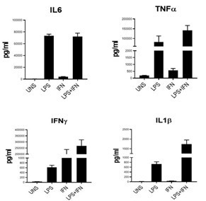
Figure 6. Cytokine release from fresh axoCells Microglia following 24-hour stimulation with LPS, INFy, or both (n = 3). UNS = unstimulated. The expected pattern of cytokine release demonstrates functional relevance. Image Credit: Axol Bioscience Ltd
Fuel high performance in assay systems
Advanced in vitro assay systems have extensively used axoCells Microglia. They have also been utilized in more than 40% of the bespoke axoServices project work, either monocultured or co-cultured with other neural cells, for investigations and compound screening initiatives.
Functional QC for axoCells Microglia
As the next stage of the quality chain, Axol has been investigating functional QC (fQC), which involves evaluating the effectiveness and performance of cells in relevant biological assays. This new standard will improve translational power in sophisticated in vitro models and boost confidence in the cells' physiological relevance.
Advanced in vitro models of Alzheimer’s disease and other neurodegenerative diseases can be powered by axoCells Microglia. A phagocytosis assay that measures bait uptake over 24 hours against a preset threshold will be part of the fQC, and it will be inhibited when cytochalasin D is added.
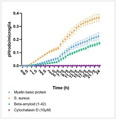
Figure 7. axoCells Microglia demonstrating phagocytosis of various baits. axoCells Microglia were thawed and matured for seven days before adding pHrodo-labeled bait. Baits were added to the cells, and phagocytosis was monitored over 24 hours using an IncuCyte® S3, showing a steady increase. Cytochalasin D (10 µM) was used as a negative control and showed complete inhibition of phagocytosis. Image Credit: Axol Bioscience Ltd
 Get the Full Guide on Human iPSC-derived MICROGLIA
Product Information
Source: Axol Bioscience Ltd
| Product Name |
Cells only code / 1 vial |
Quantity* / per vial |
Kit** code |
axoCells™ Human iPSC-Derived Microglia,
male donor, ≥1 million cells |
ax0664 |
≥1 million cells |
ax0679 |
axoCells™ - Neurons and neuroinflammatory cells