Osteoblasts are the cells required for bone synthesis and mineralization, both during the initial formation of bone and during bone remodelling.
These cells are present on the bone surface in the form of a closely packed layer, from which processes extend from the osteoblast body through the developing bone.

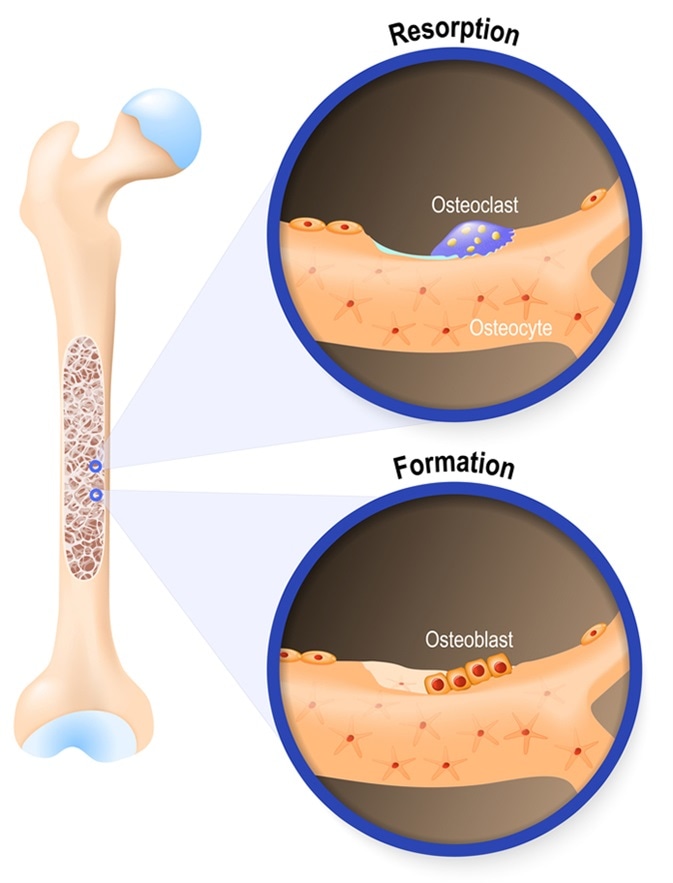
Osteoblast and osteoclast. The bone remodeling process. In a healthy body, osteoclasts and osteoblasts work together to maintain the balance between bone loss and bone formation. Image Credit: Designua / Shutterstock
These bone-forming cells are formed when osteogenic cells differentiate in a tissue covering the outer surface of bone, called the periosteum. They also arise from osteogenic cell differentiation occurring in the endosteum, a structure found in the middle of bone and in the bone marrow.
Osteoblasts produce many substances including growth factors, collagenase, osteocalcin, alkaline phosphatase and collagen.
The bone matrix eventually grows to surround osteoblasts and the bone material becomes calcified. Once surrounded and trapped, the osteoblast becomes a mature bone cell referred to as an osteocyte.
Osteoblasts and Osteoclasts
Bone Formation
The development and growth of bone is referred to as osteogenesis or ossification. Some skeletal bone begins to form during the first few weeks after conception and after eight weeks, cartilage and connective tissue have developed a skeletal pattern, at which point ossification starts.
The bone continues to develop throughout adulthood to repair fractures and remodel bone.
There are two types of bone formation or ossification, namely, intramembranous ossification and endochondral ossification.
Intramembranous Ossification
In this process, the connective tissue membranes are replaced with bony tissue to form bones referred to as intramembranous bones. Examples of bone formed in this way are the skull, the mandible and the clavicles.
Osteoblasts migrate to the connective tissue membranes where they deposit bony matrix that then surrounds them, at which point they become osteocytes.
Here, hyaline cartilage is replaced with bony tissue, which is how the majority of skeletal bones are formed. Bones that form in this way are referred to as endochondral bones.
During the end of the first trimester, osteoblasts and blood vessels infiltrate the perichondrium surrounding the hyaline cartilage, at which point the perichondrium becomes the periosteum.
The osteoblasts create a band of compact bone that surrounds the mid-section of bone called the diaphysis, while cartilage in the middle of this structure starts to break down and be penetrated by osteoblasts that replace the cartilage with spongy bone, to create a primary ossification center.
This ossification extends outwards from the center and continues to the bone ends called epiphyses.
Within the end bones, the cartilage carries on growing, which increases the length of the bone as it develops. Following birth, secondary ossification centers start to form in these bone ends.
Once the centers are formed, bone replaces the hyaline cartilage in all but two regions – the surface of the bone end stays coated in hyaline cartilage and articular cartilage and stays in place between the bone end and the diaphysis.
At the epiphysial growth plate, bones increase in length in a similar way to how endochondral ossification occurs.
Cartilage in part of the plate adjacent to the bone end undergoes mitosis and proceeds to grow. In the area adjacent to the diaphysis, cells in the cartilage called chondrocytes begin to age and degrade.
Osteoblasts migrate to the area and the matrix becomes ossified, forming bone. This is an ongoing process during childhood and into early adulthood when growth of the cartilage slows down and eventually ceases.
This generally happens in the early twenties, at which point the epiphyseal growth plate undergoes complete ossification and no further bone growth occurs.
Although the bones stop increasing in length during early adulthood, bone can still thicken in response to factors such as weight gain or increased muscle activity, a process referred to as appositional growth.
Bone diameter is increased as osteoblasts create compact bone that surrounds the outer surface of the bone and osteoclasts break down bone on the inner surface. This enables the bone to thicken, without it becoming too large and heavy.
Further Reading
Last Updated: Feb 27, 2019