Market leaders in temperature controlled microscopy, Linkam Scientific Instruments report on the use of their innovative CMS196 cryo stage for the study of mammalian cells at the London Research Institute, Cancer Research UK.
 For mammalian cells to remain in a healthy state, they require constant renewal of their components. The process of disposing of old components is known as 'autophagy', which stems from the Greek words auto "self" and phagein "eat". This process involves the formation of a double-membrane structure called an autophagosome, which engulfs old or dysfunctional organelles and then fuses with lysosomes, where they are broken down to recycle the constituent molecules. Autophagy is increased when cells are starved, and plays a fundamental role in a large number of cellular processes, including development, immunity, neurodegeneration and cancer.
In a recent publication in the journal, Ultramicroscopy (Duke et al., 2013), Dr Lucy Collinson (LRI Electron Microscopy Unit), in collaboration with Dr Sharon Tooze (LRI Secretory Pathways Lab), imaged forming autophagosomes in whole mammalian cells. The structures are particularly difficult to capture in cells prepared for electron microscopy, so they are now using a powerful new technique called cryo-soft X-ray tomography, cryo-SXT, working with Dr Liz Duke at the Diamond Light Source synchrotron. This allows whole mammalian cells to be imaged as close to the living state as possible. The cells are grown on tiny gold grids and plunged into liquid ethane to preserve the cells in the frozen state.
In order to find the autophagosomes within the cells, they are labelled with green fluorescent protein (GFP). The fluorescent autophagosomes are then located using a technique called correlative cryo-fluorescence and cryo-soft x-ray microscopy (cryo-CLXM). Cryo-fluorescence microscopy is performed using the Linkam CMS196 stage prior to the cells being transported in cryo-containers to synchrotrons in Oxfordshire, Berlin and Barcelona for imaging. One of the major advantages of this new correlative approach is that the CMS196 stage allows the cells to be screened for quality and protein localisation in the research laboratory before actually travelling to the synchrotron, which is critical in terms of cost and efficiency. The combination of cryo-fluorescence microscopy and cryo-SXT allows scientists to link the functionality of proteins to their near native-state structure. This should find wide applications in cell biology studies of health and disease.
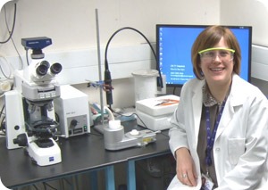
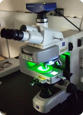
Linkam CMS196 cryo-TEM stage at the London Research Centre of Cancer Research UK with principal user, Dr Lucy Collinson.
The Linkam CMS196 stage was designed specifically to solve the problem of how to get vitrified EM grids from the fluorescence microscope into the cryo-TEM without devitrification and contamination through condensation. The stage has been optimized optically to enable the use of high NA lenses. Up to 3 grids can be loaded into a specially designed cassette for transportation from the plunge freezer to the upright fluorescent microscope. The cassette is then easily loaded onto the viewing bridge using special manipulation tools. The sample viewing chamber is perfectly dry and below -180ºC while the sample bridge itself is at -196°C. The grids can be quickly and efficiently scanned using a 100X 0.75NA lens and manipulated using high precision micrometers. The cassette is then simply manipulated back into the transportation device and is then transported to the cryo-TEM under liquid nitrogen.
Visit Linkam at www.linkam.co.uk and learn about the broad range of applications in the field of temperature-controlled microscopy.
 Linkam stages used to study the effects of cryopreservation on the quality of Koala spermatozoa samples