Leica Microsystems will be revealing a new generation of augmented reality imaging technologies for surgical microscopes, at the American Association of Neurological Surgeons (AANS) Annual Scientific Meeting 2017, in Los Angeles, USA.
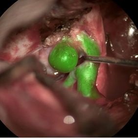
GLOW800 augmented reality fluorescence integrated with a M530 OH6 neurosurgical microscope. Photo courtesy of Cleopatra Charalampaki, MD, PhD, Professor of Neurosurgery, Department of Neurosurgery, Cologne Medical Center, Germany
Augmented reality imaging technologies supplement the microscope view, in order to support surgical decision-making and teaching during intricate neurosurgical procedures. Visitors to booth #1939 will be among the first to experience a groundbreaking new approach to vascular fluorescence during neurosurgery, known as GLOW800 augmented reality fluorescence. Further technologies being showcased at AANS include the new CaptiView HD image injection, 3D visualization, and the unique capability of the OH microscope family from Leica to display three types of intrasurgical fluorescence.
“Leica Microsystems is and always has been committed to developing innovative new ways to support its customer’s needs and requirements. In delicate neurosurgery the outstanding optical quality provided by our microscopes is not always enough. This customer pain point motivated us to develop innovative new ways to augment surgical visualization so that surgeons can operate with as much knowledge and confidence as possible,” says Markus Lusser, President Leica Microsystems. “Moreover, we ensure these new augmented technologies can be fully integrated into our premium neurosurgical microscope at any time. This gives surgeons and hospitals complete flexibility, protects their investment, and helps them remain at the cutting edge when we introduce the next advance in augmented reality imaging.”
GLOW800 augmented reality fluorescence will combine the high contrast of Near Infrared (NIR) fluorescence imaging with the full visual spectrum of white light into a single, real-time image. Instead of today’s cumbersome workflow of switching between the white light microscope view and a black and white fluorescence image, the surgeons will be able to see the white light image combined with colored NIR fluorescence flow in real-time. By overlaying the colored AR fluorescence image in the oculars, the surgeon has a more complete view of anatomical structures with no interruption or reorientation needed.
The CaptiView HD image injection system allows the surgeon to view full high-definition (HD) image data from image-guided surgery (IGS) systems and vascular fluorescence directly in the eyepieces of a Leica microscope. Without image injection neurosurgeons need to look away from the surgical field at a screen. With CaptiView image injection they can work without interruption, with the confidence that they have the high-resolution, and high-contrast data needed to make informed treatment decisions.
The American Association of Neurological Surgeons (AANS) Annual Scientific Meeting 2017, takes place in Los Angeles, USA from April 24 to April 26. “We are excited to place our new augmented reality imaging solutions in the hands of our customers at AANS,” Lusser says. “In combination with the outstanding optical quality of our M530 microscope platform, surgeons will have a wealth of visual data that will help them to achieve their goal of providing the best possible outcomes during neurosurgery.”
 Revolutionizing the fluorescence microscopy landscape with the all-in-one Microhub, Mica