Pharmaceutical research involves creation of new drugs or continuous improvement of existing drugs. This versatile research topic is a broad field of study dealing with an increasing number of challenges, owing to new pathogens emerging constantly and known pathogens becoming increasingly resistant to existing drugs.
Since more information is needed, the use of advanced tools, such as scanning electron microscopes (SEMs), has been shown to be very powerful in various applications in the pharmaceutical field. In pharmaceutical research, SEMs are used for powder imaging and analysis, to gain insights into cellular interactions with new drugs, and for applications in the most complicated cancer treatments.
This article discusses a few examples to illustrate the successful application of SEMs in research facilities across the world to develop novel and more powerful drugs to treat diseases.
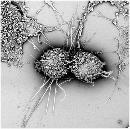
Figure 1. An example of mammalian cells observed with SEM
Superporous hydrogels
A research team at AIMST University in Malaysia is involved in the development of a new class of superporous hydrogel beads. Hydrogels consist of a network structure of cross-linked hydrophilic polymers that are capable of absorbing water in large amounts without dissolving.
These beads are employed as carriers in pharmaceuticals for controlled drug delivery based on their biodegradation and swelling abilities. Since a targeted drug delivery eliminates side effects on other cells or tissues, it helps achieve easier and faster regeneration.
With developments in research, and the increasing need to develop enhanced and highly performing drugs, the researchers have developed a new class of hydrogels with rapid swelling capacities - the reason for the name “Superporous Hydrogels”. Here, the researchers examined the surface structure and porosity of the dried beads using a SEM.
Cancer drug research
A new kind of approach was used in cancer drug research. It was found that aromatase, an enzyme responsible for determining the final sex phenotype of fish, was also playing a key role in the progression of breast cancer. Therefore, in a study, researchers used aromatase inhibitors for breast cancer treatment.
In fish, androgens are irreversibly converted into estrogens by aromatase enzymes, thereby establishing the embryo’s gender to female. Researchers are showing more interest to study male fish than female fish of this species because male fish generate anti-aromatase in large quantities.
Like Nile tilapia which gained attention as a source of food worldwide, aromatase obtained from this fish was the subject of interest for researchers looking for aromatase inhibitors.
Nile tilapia microsomes were used to study the enzyme activity of aromatase inhibitors. They are vesicle-like fractions of the endoplasmic reticulum (ER) present in healthy living cells. In this study, hepatic microsomes were prepared from Nile tilapia and their morphology was explored using a SEM.
The proliferation of cancer cells was investigated using HepG2 human hepatoma cells and MCF-7 human breast cancer cells. The study results revealed that the growth of both cancer cell lines was efficiently inhibited by a specific anti-aromatase present in microsomes.
For a successful cancer research, the morphology of tissues needs to be analyzed and understood. At present, this can be achieved using the correlated light and electron microscopy technique.
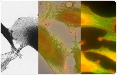
Figure 2. Correlated light and electron microscopy image of HeLa cells
Development of antibacterial powders
Antibiotics are excessively used and as a result, the numbers of antibiotic resistant bacteria are constantly increasing. Hence, researchers are seeking new ways to find the presence of bacteria on medical devices to prevent nosocomial infections or hospital-acquired infections as much as possible. Extensive research is going on in the development of new antibacterial powders.
A study revealed that pathogens present on polymer medical appliances can be very efficiently destroyed when ZnO and Ag-ZnO crystals are added to antibiotics. Here, a SEM was used to analyze the elemental composition and morphology of the crystals before using them for further experiments.
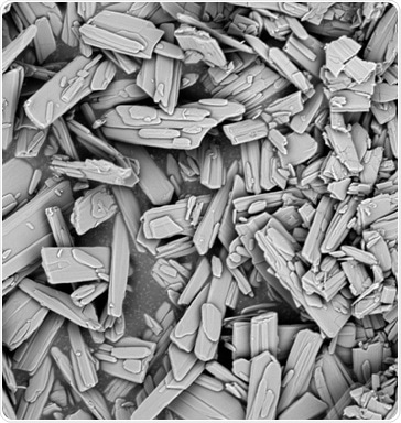
Figure 3. Pharmaceutical crystals observed with SEM
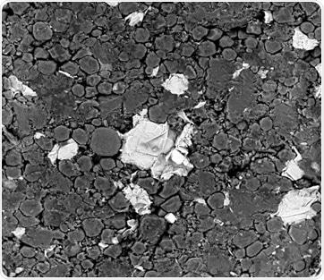
Figure 4. Pharmaceutical components including crystals observed with SEM
Conclusion
In summary, scanning electron microscopes are a very polyvalent tool that can be used for various research activities in the pharmaceutical field. It helps understand the morphology of the component of interest and highlights the effect of interactions with its environment.
References
- Development and in vitro Evaluation of New Generation Superporous Hydrogel Beads (SPHBs) Containing Fluconazol. Kumar et al. Journal of Pharmaceutical Sciences & Research; Vol. 5 Issue 12, p259 (2013)
- Investigation of anti-aromatase activity using hepatic microsomes of Nile tilapia (Oreochromis niloticus). Pikulkaew et al., Drug Discoveries and Therapeutics (2017)
- Antibacterial Powders for Medical Application Prepared by Microwave Hydrothermal Assisted Synthesis. Kunitka et al,. Nanoscience and Nanotechnology, 6(1A): 88-91 (2016)
About Phenom-World

Phenom-World believe breakthroughs happen when complex nanotechnology is made intuitive, easier to use and brought within reach. As the leading global supplier of desktop scanning electron microscopes, their aim is to make imaging and analysis at the nanoscale available to every scientist in every lab.
That’s why Phenom-World invest their time and effort into developing high-quality electron microscope solutions that are functionally rich, yet simple to use. Because their mission is to create technology that has a real impact on how people work.
This takes innovative thinking, collaborative working and an entrepreneurial attitude. At their home in the high-tech region of Eindhoven in The Netherlands, Phenom-World have a group of passionate people who are given the freedom they need to innovate. Plus a highly skilled team of specialists in more than 40 countries who can provide support on a global scale.
Cooperation and co-creation are key to Phenom-World's success. They work closely with their partners to ensure they have the latest technologies, state-of-the-art equipment and applications for imaging and analysis. So they can get fast and accurate results and achieve more in nanotechnology.
Phenom-World is globally the yearly number 1 manufacturer of desktop scanning electron microscopes and imaging and analysis packages.
Sponsored Content Policy: News-Medical.net publishes articles and related content that may be derived from sources where we have existing commercial relationships, provided such content adds value to the core editorial ethos of News-Medical.Net which is to educate and inform site visitors interested in medical research, science, medical devices and treatments.