Researchers find solutions to the issues that are faced daily in medicine by combining biology with mathematics, physics, or chemistry.
These research fields address various problems through the development of bionic parts, implants, medical devices and imaging equipment, along with tissue engineering. It involves extremely delicate and very detailed work that requires very high precision.
Scanning electron microscopy (SEM) imaging is a robust tool to study such details at the nanoscale and to push the analyzes further. SEMs are highly versatile instruments that are shown to be particularly useful in various biomedical research fields, ranging from orthodontics to bone grafts and tissue engineering.
Applications of SEM
It is a big challenge to treat bone lesions caused by tumors, diseases, or trauma. Hence, Bone lesion treatment is a key subject of interest in orthopedic research. In a 2016 study led by a laboratory in Germany, bone lesion treatment with allografts1, which are tissues obtained from the patient’s own body, was compared with the treatment involving synthetic or processed xenogeneic bone grafts, which are tissues obtained from another person.
The laboratory used a SEM for this comparison, wherein the growth of human osteoblast (bone matrix cells) on BioOssⓇ, a human-like synthetic bone matrix, and on allogeneic bones after 1 week and then 3 weeks of culture, were compared. These images help concluding that the performance of the two materials is equal and confirm the efficiency of highly-processed synthetic grafts.
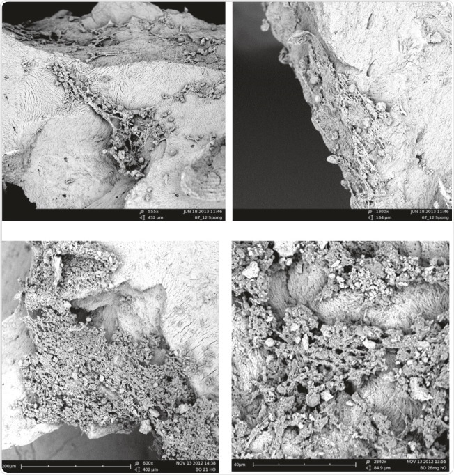
Figure 1. Top row: examples of SEM of human osteoblasts seeded on cancellous bone (ACB) after 1 week of cultivation, showing embedded cells in a bone typical extracellular matrix (image magnification 555x and 1300x as indicated). Lower row: After 3 weeks of cultivation an extensive growth of the osteoblasts could be observed on the surface on BioOss® (image magnification 600x and 2840x as indicated). Images adapted1.
In another study on skeletal injuries and tissue engineering conducted in Poland, the application of titanium scaffolds in tissue engineering2 was evaluated. It is possible to replace bone loss using tailored titanium scaffolds, which are supporting net shaped structures, created by additive manufacturing. It is essential to remove these additives during a chemical treatment used for polishing the scaffolds.
The effect of this chemical polishing on the porosity and morphology of the scaffolds was studied using a SEM. The size of the chemical particles was determined using the ParticleMetric software and the morphology before and after polishing was shown by the subsequent SEM imaging. The study was successfully completed using the PoroMetric software using which the effect of chemical polishing on the scaffold’s pores was visualized and measured by the researchers.
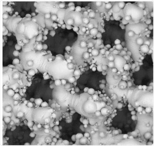
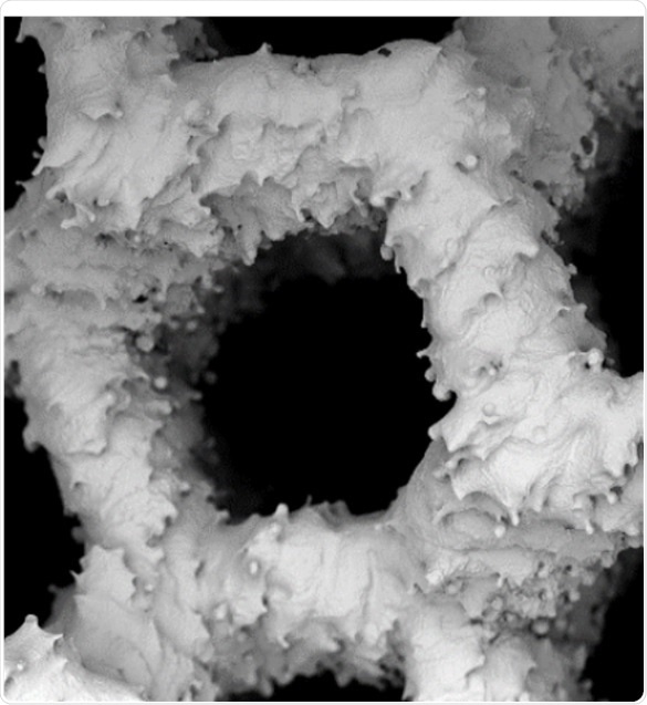
Figure 2. SEM images of scaffold. Adapted from 1.
A team of Australian researchers3 adopted a different method to study the impact of salivary pH on orthodontic stainless steel arch-wires. The objective of this study was to link corrosion with salivary pH variations and the arch-wire bending properties of stainless steel.
The SEM made it possible to establish surface irregularities owing to corrosion at varying pH levels. Subsequently, it was established that the stainless steel arch-wire should be bent carefully, especially with patients whose salivary pH is unstable, because the arch-wire tends to corrode more quickly.

Figure 3. Representative SEM images of SS wire of control group, pH 5.6, 6.6, and 7.6, respectively.
With these three instances, this article has demonstrated the range of biomedical applications that can be achieved with SEM.
References
1 Clinical Trial and in vitro study Comparing the Efficacy of Treating Bony Lesions with Allografts Versus Synthetic or Highly-Processed Xenogeneic Bone Grafts. Kubosch et al. BMC Musculoskeletal Disorders (2016)
2 Post Processing and Biological Evaluation of the Titanium Scaffolds for Bone Tissue Engineering. Wysocki et al. Materials (2016)
3 Synergistic Effect of Wire Bending and Salivary pH on Surface Properties and Mechanical Properties of Orthodontic Stainless Steel Arch-Wires. Hobbelink et al. Progress in Orthodontics (2015)

This information has been sourced, reviewed and adapted from materials provided by Phenom-World BV.
For more information on this source, please visit Phenom-World BV.