Image Credit: Shutterstock/ Kateryna Kon
“The pipeline we developed is time‐efficient, reproducible, and generates high‐density quantitative information”. Rallapalli H,
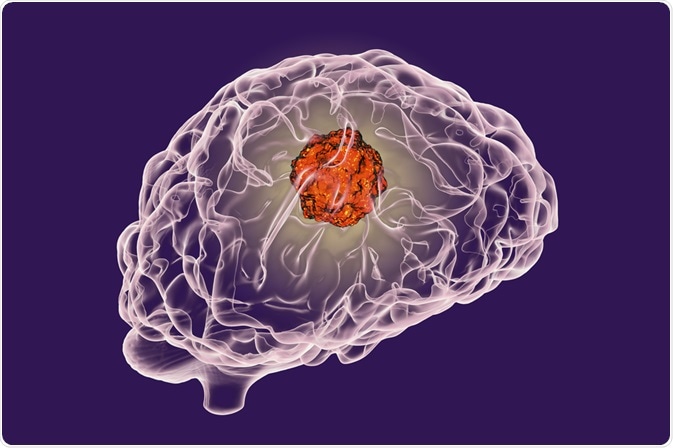
Medulloblastoma is a rare malignant brain tumor originating in the cerebellum. It is the most common cancerous brain tumor affecting children. Although treatment with surgery, chemotherapy, and radiation therapy has a high success rate, survivors commonly suffer severe treatment-related cognitive and neurological disorders.
There is therefore a need for novel targeted therapeutics for medulloblastoma that has fewer off‐target effects. To facilitate the development of such treatments, animal models have been created for studying tumor biology and testing compounds with the potential for treating medulloblastoma. Animal models have played an invaluable role in the unraveling of important concepts in physiology and in the development of novel treatment approaches for numerous disease processes.
There are four distinct molecular subgroups of medulloblastoma, and each has its own characteristic signaling pathways, activating mutations, and cell types of origin. Mouse models have been created for each of the four types of medulloblastoma. These have been genetically engineered so that the natural tumor microenvironment is maintained along with the heterogeneity and clonal nature of tumor growth.
Since medulloblastoma typically occurs sporadically, inducible genetic systems have been used in the animal models of medulloblastoma. For example, a variant of the oestrogen receptor that is only activated in the presence of tamoxifen may be used. This enables researchers to initiate medulloblastoma formation in small numbers of cells of origin at a desired developmental time‐point.
Tumor growth and response to treatment is monitored non-invasively using in vivo imaging techniques. The method of choice for investigation is Magnetic Resonance Imaging (MRI). This technique offers a high spatial resolution for resolving small tissue features in early-stage tumors. It additionally has the added benefit of being highly reproducible and providing detailed images of the whole brain. which is important since medulloblastoma can travel in cerebrospinal fluid, as so has the potential to affect other areas of the brain distant from the area of origin.
The efficiency of MRI in studying medulloblastoma has recently been improved with the development of a novel manganese‐enhanced MRI‐based technique. Sporadic medulloblastoma was modelled in mice by inducing expression of an activated form of the smoothened gene (aSmo), which transduces the hedgehog signal that is thought to drive activation of target cancer genes. A Bruker Biospec was employed to image the aSmo mice using the 3D manganese‐enhanced MRI methodology at defined time‐points.
The images obtained revealed that the probable origin of the aSmo medulloblastoma tumors was extremely specific and that there was a high level of heterogeneity between tumors. Tumour heterogeneity was observed both between different tumors in the same mouse and between tumors in different mice.
The latest MRI methodology demonstrated the capacity to provide high resolution, quantitative information relating to tumour progression and response to novel therapies in mouse models of medulloblastoma in a time-efficient manner.
Rallapalli H, et al. Magn Reson Med. 2019;00:1–14.
About Bruker BioSpin Group
The Bruker BioSpin Group designs, manufactures, and distributes advanced scientific instruments based on magnetic resonance and preclinical imaging technologies. These include our industry-leading NMR and EPR spectrometers, as well as imaging systems utilizing MRI, PET, SPECT, CT, Optical and MPI modalities. The Group also offers integrated software solutions and automation tools to support digital transformation across research and quality control environments.
Bruker BioSpin’s customers in academic, government, industrial, and pharmaceutical sectors rely on these technologies to gain detailed insights into molecular structure, dynamics, and interactions. Our solutions play a key role in structural biology, drug discovery, disease research, metabolomics, and advanced materials analysis. Recent investments in lab automation, optical imaging, and contract research services further strengthen our ability to support evolving customer needs and enable scientific innovation.
Sponsored Content Policy: News-Medical.net publishes articles and related content that may be derived from sources where we have existing commercial relationships, provided such content adds value to the core editorial ethos of News-Medical.Net which is to educate and inform site visitors interested in medical research, science, medical devices and treatments.