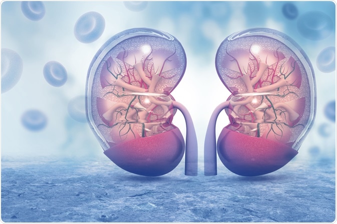
Acute kidney injury (AKI) is an episode of kidney failure which decreases the filtering capacity of the kidneys severely and happens very fast, within a few days or a few hours. Therefore, waste products that are usually filtered out by the kidneys build up in the blood, which can lead to vomiting and nausea.

Image Credit:Shutterstock/ crystal light
Furthermore, blood potassium levels rise, which can result in paralysis, muscle weakness, and heart arrhythmias. Consequently, this carries the risk of pulmonary oedema developing. AKI is associated with poor outcomes and is most common among critically ill patients.
The growth in mortality and morbidity is marked among children especially, where AKI can lengthen the time they need to be in a hospital, in intensive care, and on a ventilator. AKI can also heighten the risk of a child developing chronic kidney disease in the future1.
Over a fifth of critically ill pediatric and neonatal patients develop AKI and the incidence is growing2. AKI can affect as many as 5%3, even in non-critically ill hospitalized children and adolescents. It is vital that AKI is diagnosed as quickly as possible since it can have such a devastating impact on prognosis.
Diagnosis of acute kidney injury
Urine output measurements and serum creatinine levels are the gold standard for diagnosing AKI. Yet, these definitions of AKI have their drawbacks, not least is the fact that they only give indirect markers of decreased glomerular filtration rate.
As such, AKI may have been present for some time before it is identified by such assessments. Additionally, creatinine- and urine output assessments do not give any indication of the specific cause of the renal impairment. So, there is an ongoing unmet requirement for a more specific, earlier diagnostic test for AKI.
A number of urinary proteins have been explored as potential biomarkers for the early diagnosis and evaluation of acute kidney injury, including kidney injury molecule-1 (KIM-1), neutrophil gelatinase-associated lipocalin (NGAL), tissue inhibitor of metalloprotease-2 (TIMP-2), and insulin-like growth factor-binding protein 7 (IGFBP7)4,5.
Sadly, none of the protein biomarkers tested to date had enough specificity for kidney injury or clinical performance to support clinical implementation6.
Metabolomic studies have demonstrated great value in the identification of biomarkers that permit diagnosis plus the prediction of patient risk and clinical outcome. This strategy is now being applied to AKI.
Metabolomic analysis in acute kidney injury
Nuclear magnetic resonance spectroscopy (NMR) enables the simultaneous analysis of all the small molecules present in a sample with minimal sample preparation. This analysis of body fluids, including urine and blood, is known as metabolomics and can supply an accurate metabolic profile, which can help with disease diagnosis, prognosis, and treatment decisions.
A functional fingerprint of the body’s pathophysiological and physiological state is supplied via untargeted metabolomics. The study of these metabolite patterns permits the identification of metabolites specific to a particular disease state.
This research has been applied across a variety of renal disorders, like diabetic nephropathy, chronic kidney disease and polycystic kidney diseases7,8. Metabolic studies have also been utilized to identify biomarkers for AKI with numerous aetiologies9,10.
Yet, few clinical studies have assessed the application of metabolomic analysis in pediatric AKI, despite the wide application of metabolomic profiling to the classification and differentiation of renal disorders.
Identifying biomarkers for pediatric kidney injury
A recent pilot study examined whether 1H-NMR urine metabolomic fingerprints and biomarkers could be utilized to differentiate separate subtypes of AKI in pediatric and neonatal patients11.
Urine samples from 31 critically ill children without AKI, 65 neonates and pediatric patients with established AKI, and 53 healthy controls were analyzed by 1H NMR by utilizing a Bruker 600 MHz Avance II spectrometer with a double resonance 5 mm BBI probe.
A comparison of the 1H NMR spectra which was gathered showed four panels of metabolites indicative of a diagnosis of AKI11. Noticeable differences in metabolite profiles amongst the patients with AKI were significantly decreased urinary citrate levels, and higher levels of valine and leucine.
In patients with AKI, concentrations of bile acid and a number of unknown compounds were also changed. In addition, these preliminary data indicated that differentiation of subtypes of AKI with different aetiologies could be possible from 1H-NMR metabolomic fingerprints.
Since the differences from healthy patients were not significant, the examination also eliminated some metabolites which had been identified as potential biomarkers for AKI in animal studies (creatinine, hippurate, indoxyl sulfate). It also showed that the metabolic changes in AKI are similar for pediatric and neonatal patients, in contrast to previous hypotheses.
This latest data shows that metabolomic differentiation of AKI in pediatric and neonatal patients is feasible according to the underlying causes.
It is hoped that the differences observed in metabolite profiles could facilitate the development of a novel method for the early identification of patients with AKI and enhance patient outcomes by allowing physicians to implement medical treatment sooner. Yet, these promising results must first be validated in larger studies.
References
- Mammen C, et al. Am. J. Kidney Dis. 2012;59:523–530.
- Kaddourah A, et al. N. Engl. J. Med. 2017;376:11–20.
- McGregor T, et al. Am. J. Kidney Dis. 2016;67:384–390.
- Malhotra R, Siew ED. Clin. J. Am. Soc. Nephrol. 2017;12:149–173.
- Waldherr S, et al. Pediatr. Res. 2019;85:678–686.
- van Duijl TT, et al. Clin. Biochem. Rev. 2019;40:79–97.
- Abbiss H, et al. Metabolites 2019;9:34.
- Chihanga T, et al. Am. J. Physiol. Renal. Physiol. 2018;314:F154–F166.
- Izquierdo-Garcia JL, et al. Am. J. Physiol. Renal. Physiol. 2019, 316, F54–F62.
- Archdekin B, et al. Pediatr. Transplant. 2019, 23, e13364.
- Muhle-Goll C, et al. Int. J. Mol. Sci. 2020;21:1187.
https://www.mdpi.com/1422-0067/21/4/1187
About Bruker BioSpin Group
The Bruker BioSpin Group designs, manufactures, and distributes advanced scientific instruments based on magnetic resonance and preclinical imaging technologies. These include our industry-leading NMR and EPR spectrometers, as well as imaging systems utilizing MRI, PET, SPECT, CT, Optical and MPI modalities. The Group also offers integrated software solutions and automation tools to support digital transformation across research and quality control environments.
Bruker BioSpin’s customers in academic, government, industrial, and pharmaceutical sectors rely on these technologies to gain detailed insights into molecular structure, dynamics, and interactions. Our solutions play a key role in structural biology, drug discovery, disease research, metabolomics, and advanced materials analysis. Recent investments in lab automation, optical imaging, and contract research services further strengthen our ability to support evolving customer needs and enable scientific innovation.
Sponsored Content Policy: News-Medical.net publishes articles and related content that may be derived from sources where we have existing commercial relationships, provided such content adds value to the core editorial ethos of News-Medical.Net which is to educate and inform site visitors interested in medical research, science, medical devices and treatments.