Dental burs are generally applied to rid teeth of residual adhesive after the removal of orthodontic brackets. During the process, these burs sustain wear degradation. The question arises, should these burs be single-use only or can they be used on another patient?
Employing micro-CT for nondestructive 3D imaging of used burs helps address this question.
As numerous teenagers and parents know, current orthodontic treatment involves aligning and moving teeth by utilizing adhesively attached orthodontic appliances (Fig. 1A). To bond orthodontic brackets to the outer labial tooth surfaces, dental composites and resins are typically used (Fig. 1B).
Upon completion of the treatment, perhaps months or years later, the brackets and the dental materials must be removed (Fig. 1C).
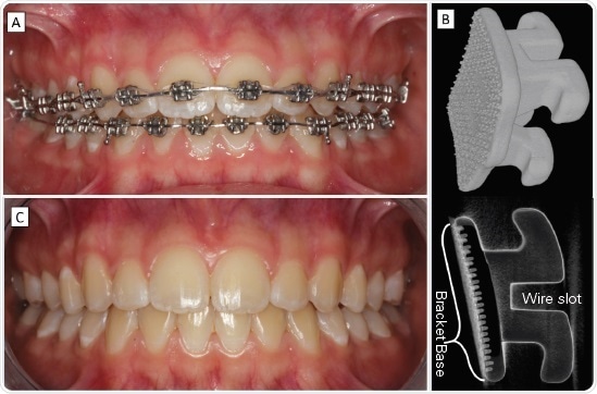
Figure 1. To align teeth (A) orthodontists use brackets (B) and wires that apply forces to change tooth position and angle. Treatment ends (C) with bracket removal, when the teeth and dental arches have reached aesthetic, harmonious well-matching relations leading to functional occlusion and pleasing smile. Image Credit: Bruker BioSpin - NMR, EPR and Imaging
Removal of the adhesive residue is a crucial part of treatment and a clinical challenge for the treating orthodontist; the adhesive must be completely removed from the tooth without resulting in any damage to the teeth.
High-velocity water-cooled burs are frequently used, which require precision, patience and complete patient cooperation. A range of grinding tools are available for this task, for instance, tungsten carbide burs (Fig. 2A, B).
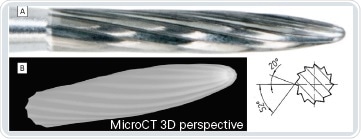
Figure 2. These example burs are designed to gently remove polymer and composite adhering to the outer tooth surface. Although informative, the optical images (A) provide only partial information about the 3D design of the tool with little information on how it operates. Tomographic imaging (B) helps to better understand its mode of action. Image Credit: Bruker BioSpin - NMR, EPR and Imaging
However, there are questions surrounding the optimal use of these tools and how best to use them.
Material and methods
Figure 3 illustrates the steps typically involved in post-orthodontic adhesive removal, utilizing the flame-shaped H48L tungsten carbide bur (Komet Dental, Gebr. Brasseler GmbH & Ko. KG, Lemgo, Germany).
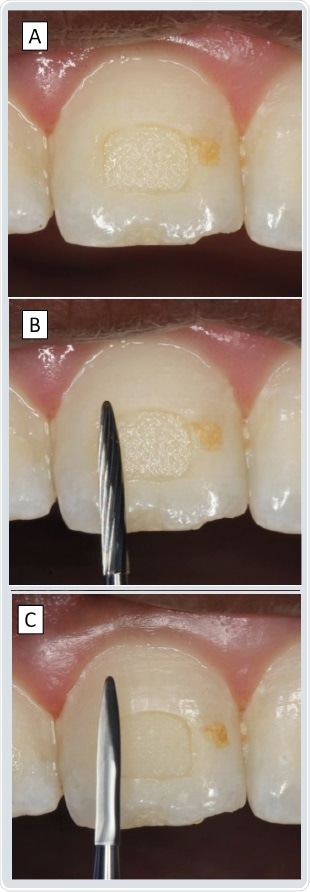
Figure 3. These example burs are designed to gently remove polymer and composite adhering to the outer tooth surface. Although informative, the optical images (A) provide only partial information about the 3D design of the tool with little information on how it operates. Tomographic imaging (B) helps to better understand its mode of action. Image Credit: Bruker BioSpin - NMR, EPR and Imaging
The tapered flutes (Fig. 2) of the bur have an angle of 52° in the direction of rotation and a modest quasi-orthogonal angle of 20° in relation to the shaft axis. This limits any potential damage to the surface of the teeth if/when the bur encounters enamel.
This particular bur has 12 flutes and a diameter of 0.14 mm and comes with various shafts developed to fit all available handpieces on the market.
Typical clinical use of the bur includes:
- Removal of the brackets with pliers; detachment commonly occurs in the bracket-adhesive interface (Fig. 2B)
- Grinding away of the composite residues from the surface of the tooth by consistently moving the water-cooled rotating bur across the polymer on the outer surface. Gentle pressure must be applied by the clinician must, facilitating appropriate timescales for the bur to remove the adhesive
- Now and then, compressed air is used to dry the surfaces and locate any remaining contaminated tooth surface patches
- Slow-speed rubber cups are used for polishing the tooth surface, which removes residues
- Application of fluoride is often demonstrated to enhance enamel resistance to erosion or bacterial contamination
To closely evaluate the wear of the bur, micro-CT was utilized to record the 3D shape and details of the geometry of flute edges, with the high-resolution settings of a desktop instrument.

Figure 4. Economic considerations in choosing bur use, characteristics and contribution to clinical workflow. Image Credit: Bruker BioSpin - NMR, EPR and Imaging
The bur was imaged using the Bruker SkyScan 1275 micro-CT (Bruker micro-CT, Kontich, Belgium) fixed to a thin sample holder enabling close positioning to the X-ray source.
The high density of elements in the bur required employing the higher energy source settings of 100 kV (yielding µA) and a CU filter helped eliminate lower energies reducing substantial edge artefacts.
Optimal scans were acquired using 360° rotations, 6 µm effective pixel size. To reconstruct the image volumes, the NRecon (V. 1.7.4.2, Bruker micro-CT, Kontich, Belgium) was used.
When using the CTvox (V3.0, Bruker micro-CT, Kontich, Belgium), 3D images may be produced instantly, the reconstructed data in slices is immediately available as TIF or PNG files to be evaluated in any other package.
Examples of conventional cross-sectional slices can be acquired using the open-source ImageJ packages (https://imagej.net, V 1.52n) and its derivative, Fiji.
Stacks of images mean it is relatively straightforward to select, compare and measure alterations in shape at the same height in the sample both before and after use. Thus, it is possible to review bur abrasion associated with usage.
Direct comparison of the bur scans before and after the bracket removal session highlights selective zones with fine but significant regions of wear (Fig. 5).
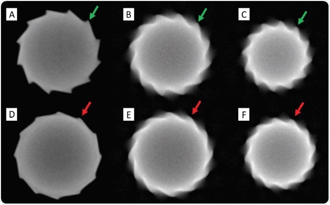
Figure 5. Dental carbide wear in 2D sections along the bur axis (H48L tungsten carbide, Komet Dental, Gebr. Brasseler GmbH & Co. KG, Lemgo, Germany): Whereas obvious rounding of the edges is seen near the shaft (compare A with D, read/green arrows) extensive wear is observed in the central area(compare B and E, red/green arrows), with notable but less significant effects at regions ¾ up towards the tip (compare C with F, red/green arrows). Image Credit: Bruker BioSpin - NMR, EPR and Imaging
Rounding of flute edges is considered symmetric around the axis of the bur but diversified and non-uniform at various cross-sections across the length of the bur.
Although there are known properties (strength, wear resistance) relative to the materials used for bracket adhesion, the association between wear patterns during composite removal and the practitioner’s technique remains largely unknown.
Future studies will make it possible to quantify such measurements to show dimensions and locations in space that can act as key input for numerical modeling.
The nature of the reconstructions makes it well-suited for more comprehensive 3D analysis. By converting the data into binary (black and white) slices and into industry-standard STL files (export function of the inbuilt Fiji volume viewer), a thorough and detailed analysis of wear is made possible.
By using programs that have the capacity to align and compare the 3D data, there is the potential to visualize and quantify the area of wear and carbide substance loss. For example, the 3D analysis software Geomagic Control (3D Systems Inc., Rock Hill, S.C., U.S.A.) highlights the variations in the 3D deviation (mismatch, in 3D) between the original unused and worn bur (Fig. 6).

Figure 6. Superimposition and comparison of the bur volumes before and after use reveal important regionally varying differences in the amount of remaining substance (Tungsten carbide in this case). The green traces indicate a mismatch (averaging at about 130 μm in the flutes) with a clear trend for loss, as opposed to yielding or deformation of the flutes. Image Credit: Bruker BioSpin - NMR, EPR and Imaging
It can be observed that significant wear is seen in the central region of the flutes and at the bur tip. The inclination is that these are related to the adhesive properties, the geometry and hardness of the bur, as well as the clinical technique being used.
Results & conclusion
Superimposition of the virtual data acquired before and after bur use highlighted that wear appears in the central region up to two-thirds of the height of the bur. Wear never surpassed 1 mm in any direction (Fig. 6).
The flutes are significantly worn after usage and would therefore necessitate greater force and an extended treatment time to eradicate any remaining composite on the surface of the teeth, possibly leading to dangerous tooth overheating.
The evaluations from the 3D micro-CT imaging imply the recommendation that a bur should be limited to just one debonding session in a single patient. However, further comparisons with different usage protocols and bonding materials are required to investigate how general this single-use recommendation should be.
About Bruker BioSpin - NMR, EPR and Imaging

Bruker BioSpin offers the world's most comprehensive range of NMR and EPR spectroscopy and preclinical research tools. Bruker BioSpin develops, manufactures and supplies technology to research establishments, commercial enterprises and multi-national corporations across countless industries and fields of expertise.
Sponsored Content Policy: News-Medical.net publishes articles and related content that may be derived from sources where we have existing commercial relationships, provided such content adds value to the core editorial ethos of News-Medical.Net which is to educate and inform site visitors interested in medical research, science, medical devices and treatments.