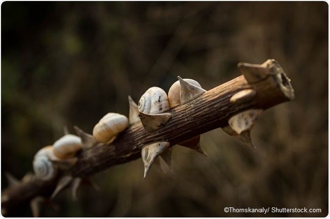
Thorn snails—relatively small, fragile Ellobioidea-related snails that are found primarily in temperate and tropical zones—represent an integral ecological component of leaf litter decomposition in stable environments.1 These small, “micro” snails mingle with insects, bacteria, and fungi to increase biodiversity in the decomposition process, ultimately assisting in the release of nutrients into the soil and improving the health of vegetation.
The study of thorn snails, including the identification of new varieties, may be imperative for not only understanding the intricacies of soil and plant development, but to also provide details regarding how researchers can take actions toward improving the environmental landscape as a whole.
The Study of Thorn Snails
To date, 14 thorn snail species have been identified throughout North and Central America. Recently, 3 new species of thorn snails have been identified using a novel approach—computed tomography (CT) imaging. The Bruker microCT system was essential for this approach due to the extreme compactness of the snails’ anatomy.
CT is widely utilized in medicine to study anatomical and physiological activity in the human body.2,3 The use of CT imaging for thorn scales requires a slightly different approach; smaller CT technology (“nano” CT) is essential for examining these creatures at the most minute level.
Dr. Adrienne Jochum et al published their findings on the application of nano-CT in micro snail identification in ZooKeeys.4 This is one of the first few studies using CT in this field. Specifically, researchers used nano-CT imaging for comparing and identifying three new Carychium species: Carychium hardiei, Carychium belizeense, and Carychium zarzaae.
Due to the snails’ anatomical features5 (they measure <2 mm in length and can often only be found by using a magnifying glass) and highly fragile structure, researchers believe CT to be the most appropriate imaging method for identification of these types of mollusks.
The discovery of these new species was entirely accidental—Dr. Jochum found one of the snails (Carychium hardiei) during a rest stop in Georgia on the way to the Atlanta Airport. The Carychium belizeense was first discovered in Belize in the Bladen Nature Reserve, and the Carychium zarzaae was discovered in Panama.
CT Scans Discover New Species of Mini Thorn Snails
In this study, the 3 newly discovered thorn snail species were analyzed under nano-CT imaging and compared to currently known species. Researchers use the Bruker microCT system as the main instrumentation. This allowed researchers to understand not only the structure of the new species, but it also enabled further identification of the snails’ potential relation to currently known snails.
Carychium hardiei
Carychium hardiei, named after American naturalist, field biologist, and chemical engineer Frank Hardie, was compared to the Carychium floridanum. The new species had a greater bulbous protoconch, a thinner peristome, and a smaller size of the deeply-set parietal denticle.
As compared with C. mexicanum under CT, it was found that the new C. hardiei had a larger, more tapered shell and also featured a smaller and thinner peristome. Following evaluation of DNA barcode data, researchers determined that the newer species substantially differed from all other known North and Central American thorn snail species.
Carychium belizeense
Carychium belizeense was found in abundant numbers and is considered common in leaf litter in the Bladen Nature Reserve. Therefore, this species is not likely to be a threatened species. Researchers were also able to gain a side view aperture-left profile of the species with the Bruker microCT.
CT imaging was also used to study the structure of the new C. belizeense, named after its country of origin Belize. Upon CT analysis, the C. belizeense species featured approximately 4 highly convex whorls (a 360° revolution in the spiral of the shell). Compared with the C. hardiei species, C. belizeense had a bigger, more robust shell and more whorl convexity.
Carychium zarzaae
Carychium zarzaae is named after Eugenia Zarza, who retrieved the material for a previous study.6 Compared with the Panamanian lineage under CT, C. zarzaae had a much thinner shell. Its peristome formation and superficial teleconoch striation resembled that of the C. belizeense and C. hardiei, respectively. Upon comparison, it was clear this snail was just as distinct as the others and represented new species of mollusks.
Micro CT in Zoology: A New Approach
As demonstrated in the findings of this study, the authors of this paper were able to gain an in-depth observation of the newly discovered snail species with CT imaging technology. Researchers in this study imaged almost all Carychium with the SkyScan 2011 nano-CT system (microCT) system by Bruker.7 The Bruker microCT system features a LaB6 cathode, transmission anode, and an “open type” [pumped] X-ray source.
For this system, researchers are not required to provide any type of coating or vacuum treatment prior to imaging. The imaging system, which includes software for sample visualization, generates 3D images of a sample’s internal microstructure and morphology at the sub-micron level. These features are essential for small samples that offer wide-range applicability in the biological, chemical, and pharmaceutical industries.
References:
- Interactions. UW-La Crosse. http://bioweb.uwlax.edu/bio210/2011/blake_zeld/Interactions.html.
- Truong QA, Hoffmann U, Singh JP. Potential uses of computed tomography for management of heart failure patients with dyssynchrony. Crit Pathw Cardiol. 2008;7(3):185-190.
- Liguori C, Frauenfelder G, Massaroni C, et al. Emerging clinical applications of computed tomography. Med Devices (Auckl). 2015;8:265-278.
- Jochum A, Weigand AM, Bochud E, et al. Three new species of Carychium O.F. Müller, 1773 from the Southeastern USA, Belize and Panama are described using computer tomography (CT) (Eupulmonata, Ellobioidea, Carychiidae). ZooKeys. 2017;675:97-127.
- Scientists discover tiny glassy snails in caves of Northern Spain. Phys.org. https://phys.org/news/2015-03-scientists-tiny-glassy-snails-caves.html.
- Weigand AM, Jochum A, Pfenninger M, Steinke D, Klussmann-Kolb A. A new approach to an old conundrum − DNA barcoding sheds new light on phenotypic plasticity and morphological stasis in microsnails (Gastropoda, Pulmonata, Carychiidae). Molecular Ecology Resources. 2011;11:255–265.
- microCT. Bruker Instruments. https://www.bruker.com/en.html
About Bruker BioSpin - NMR, EPR and Imaging

Bruker BioSpin offers the world's most comprehensive range of NMR and EPR spectroscopy and preclinical research tools. Bruker BioSpin develops, manufactures and supplies technology to research establishments, commercial enterprises and multi-national corporations across countless industries and fields of expertise.
Sponsored Content Policy: News-Medical.net publishes articles and related content that may be derived from sources where we have existing commercial relationships, provided such content adds value to the core editorial ethos of News-Medical.Net which is to educate and inform site visitors interested in medical research, science, medical devices and treatments.