Sponsored Content by MerckReviewed by Emily MageeJan 15 2024
The three-dimensional (3D) printing of biological tissue is quickly becoming a vital part of tissue engineering. Advancements in 3D printing technology have facilitated the development of many new fabrication methods, enabling the production of cell-laden constructs,1-2 composite tissue,2 and scaffold-free tissue.3 These advances pave the way for the eventual printing of whole functional organs from raw materials.
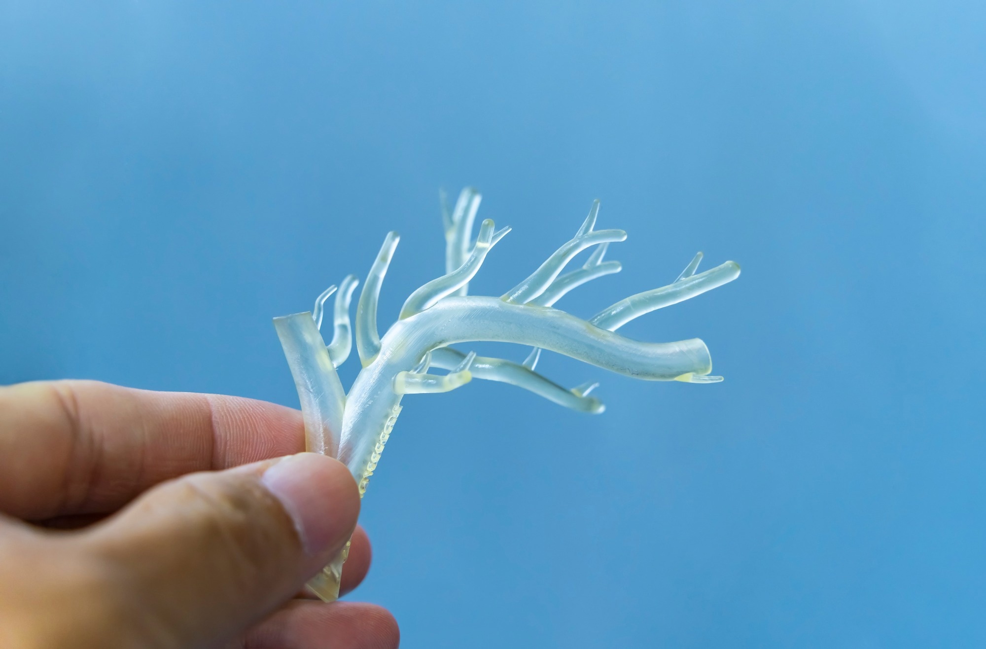
Image Credit: asharkyu/Shutterstock.com
Additionally, 3D printing can be employed to produce tissue engineering scaffolds that replicate the 3D microenvironment of the native organ. For the fabrication of tissue scaffolds, 3D printing is amenable to a variety of biomaterials, including collagen, calcium phosphate,4 hydrogel,6 polycaprolactone (PCL),2,8 CaSiO3,5 hydroxyapatite,7-8 and starch-based polymers.9
This flexibility enables the fabrication of composite structures that best replicate the physiological conditions that surround the tissue, such as mechanical properties and topographical cues.10 Due to these properties, 3D-printed scaffolds can act as desirable support structures for the 3D cultivation of cells.
However, characterizing 3D printed tissue frequently requires preparatory steps that are not employed in conventional 2D tissue culture analysis. For instance, employing light microscopy methods like confocal microscopy to image through thick 3D tissue is difficult because of scattered light in biological samples. As a result, many imaging techniques are applied to tissue sections that have a thickness of less than 20 µm.11
Although innovative imaging methods report successful visualization of much thicker tissues, they are still limited to tissues that are at most several millimeters thick.11 Therefore, proper sectioning is important for the characterization of multilayer cell constructs produced by 3D printing.
Biological stains should be carefully chosen to extract the most relevant information about the printed tissue, including cell viability and maturation. For example, determining the cell viability in the inner core of a 3D printed construct is crucial as these cells can have a higher likelihood of necrosis caused by insufficient nutrient exchange.12
This article discusses the processing and staining approaches for the evaluation of 3D-printed tissues and scaffolds.
Confirming material compatibility with cells of interest
Before fine-tuning a 3D printing method for biological use, it is important to check that the material works well with the specific cells, allowing them to stick to it and grow. This can be done by testing cell survival and growth on the material before printing.
Preparing 3D printed tissues/scaffold for light microscopy
Once a cellular construct has been successfully printed or grown on a scaffold, a chemical fixative (detailed in Table 1) is applied before any embedding or sectioning takes place. This is to prevent any change or loss of cellular components during processing.
Alternatively, tissue can be frozen and cryo-sectioned if chemical fixation is not desired.
Although the utilization of formalin solution is common in tissue fixation,13 the choice of fixative is highly dependent on the tissue’s specific properties as well as the purpose of the study.
Factors that can influence fixation include the mechanism of the fixative (e.g., denaturation or crosslinking) 13 and the condition of the fixation procedure (e.g., duration of tissue exposure to fixative and temperature).13-14
Additionally, non-toxic agents that do not contain glutaraldehyde or formaldehyde are available for tissue fixation, such as the HistoChoice® tissue fixative and clearing agent from Merck. Table 1 details available tissue processing products.
Table 1. Various fixatives used for tissue fixation. Source: Merck
| Product No. |
Description |
| 340855 |
Glutaraldehyde solution 50 wt. % in H2O |
| HT501128 |
Formalin solution, neutral buffered, 10% histological tissue fixative |
| P6148 |
Paraformaldehyde reagent grade, crystalline |
| H0290 |
Hartman′s Fixative histological tissue fixative |
| Z2902 |
Zinc Formalin Fixative |
| A5472 |
Formalin Free Tissue Fixative |
|
| 2858 |
Ethanol Fixative 80% v/v suitable for fixing solution (blood films) |
| H2904 |
HistoChoice® Tissue Fixative |
| H2779 |
HistoChoice® Clearing Agent |
After being properly fixed, the tissue can be sequentially dehydrated in ethanol and embedded in a medium like Paraplast® (detailed in Table 2) for sectioning.13 The embedded tissue may be sectioned in a desired thickness onto glass slides (as shown in Table 2) via a microtome.
The thickness of sections must be determined based on the specific type of printed tissue and the biological stains that will be utilized. For instance, while muscle tissues can be sectioned in slices that are 4-6 µm in thickness, it is recommended that brain or spinal cord samples are sectioned into thicker (10-40 µm) slices.15
Table 2. Materials used for processing and embedding tissue samples. Source: Merck
| Product No. |
Description |
| P3558 |
Paraplast® for tissue embedding |
| P3808 |
Paraplast X-TRA® for tissue embedding |
| P3683 |
Paraplast Plus® for tissue embedding |
| E7023 |
Ethyl alcohol, Pure 200 proof, for molecular biology |
|
| 534056 |
Xylenes histological grade |
| S8902 |
Slides, microscope plain, size 25 mm × 75 mm |
| S8400 |
Slides, microscope frosted one end, size 25 mm × 75 mm |
| S9027 |
Slides, microscope opaque (white), size 25 mm × 75 mm |
| C9802 |
Cover glasses size 22 mm × 22 mm |
| C7931 |
Cover glasses size 24 mm × 40 mm |
| C8181 |
Cover glasses size 24 mm × 50 mm |
| C9056 |
Cover glasses size 24 mm × 60 mm |
| P5493 |
Phosphate buffered saline 10× concentrate, BioPerformance Certified, suitable for cell culture |
Once the tissue is sectioned onto microscope slides, different staining procedures may be applied using the buffers and stains detailed in Table 3. For a specific antigen of interest, immunohistochemistry (IHC) methods may be employed.
Following IHC, samples are frequently co-stained using a counterstain, such as hematoxylin or DAPI.
Table 3. Popular biological stains and buffers. Source: Merck
| Product No. |
Description |
| D9542 |
DAPI for nucleic acid staining |
| P5282 |
Phalloidin, Fluorescein Isothiocyanate Labeled sequence Amanita phalloides(synthetic: peptide sequence) |
| P1951 |
Phalloidin–Tetramethylrhodamine B isothiocyanate sequence from Amanita phalloides(synthetic: peptide sequence) |
| T6146 |
Trypan Blue powder, BioReagent, suitable for cell culture |
| T8154 |
Trypan Blue solution 0.4%, liquid, sterile-filtered, suitable for cell culture |
| 81845 |
Propidium iodide ≥94% (HPLC) |
| P4864 |
Propidium iodide solution solution (1.0 mg/ml in water) |
| HHS16 |
Harris Hematoxylin Solution, Modified |
| HHS32 |
Harris Hematoxylin Solution, Modified |
| HHS80 |
Harris Hematoxylin Solution, Modified |
| HHS128 |
Harris Hematoxylin Solution, Modified |
| P5493 |
Phosphate buffered saline 10× concentrate, BioPerformance Certified, suitable for cell culture |
| A9647 |
Bovine Serum Albumin heat shock fraction, pH 7, ≥98% |
| P3688 |
Phosphate buffered saline pH 7.4, contains BSA, powder |
| P9416 |
TWEEN® 20 for molecular biology, viscous liquid |
| T8787 |
Triton™ X-100 for molecular biology |
Imaging 3D printed tissue/scaffold using scanning electron microscopy (SEM)
In addition to the above-mentioned light microscopy methods, SEM analysis may be required for the visualization of the 3D printed construct’s nanoscale morphology. SEM imaging is beneficial for 3D-printed scaffolds that are created to replicate the micro/nanostructures of the tissue microenvironment.
For SEM preparation, biological samples undergo a series of steps. First, they are fixed using EM grade fixatives (refer to Table 4), followed by sequential dehydration with ethanol. Next, they are subjected to critical point drying. Finally, the samples are coated with a conductive material, such as gold, to minimize charging artifacts.
Table 4. Chemicals used for fixation and dehydration for SEM imaging preparation. Source: Merck
| Product No. |
Description |
| G7776 |
Glutaraldehyde solution Grade I, 70% in H2O, specially purified for use as an electron microscopy fixative or other sophisticated use |
| G7651 |
Glutaraldehyde solution Grade I, 50% in H2O, specially purified for use as an electron microscopy fixative or other sophisticated use |
| G5882 |
Glutaraldehyde solution Grade I, 25% in H2O, specially purified for use as an electron microscopy fixative |
| G7526 |
Glutaraldehyde solution Grade I, 8% in H2O, specially purified for use as an electron microscopy fixative or other sophisticated use |
| E7023 |
Ethyl alcohol, Pure 200 proof, for molecular biology |
References
- Kolesky DB, Truby RL, Gladman AS, Busbee TA, Homan KA, Lewis JA. 2014. 3D Bioprinting of Vascularized, Heterogeneous Cell-Laden Tissue Constructs. Adv. Mater.. 26(19):3124-3130. https://doi.org/10.1002/adma.201305506
- Lee J, Hong JM, Jung JW, Shim J, Oh J, Cho D. 3D printing of composite tissue with complex shape applied to ear regeneration. Biofabrication. 6(2):024103. https://doi.org/10.1088/1758-5082/6/2/024103
- Norotte C, Marga FS, Niklason LE, Forgacs G. 2009. Scaffold-free vascular tissue engineering using bioprinting. Biomaterials. 30(30):5910-5917. https://doi.org/10.1016/j.biomaterials.2009.06.034
- Inzana JA, Olvera D, Fuller SM, Kelly JP, Graeve OA, Schwarz EM, Kates SL, Awad HA. 2014. 3D printing of composite calcium phosphate and collagen scaffolds for bone regeneration. Biomaterials. 35(13):4026-4034. https://doi.org/10.1016/j.biomaterials.2014.01.064
- Wu C, Fan W, Zhou Y, Luo Y, Gelinsky M, Chang J, Xiao Y. 2012. 3D-printing of highly uniform CaSiO3 ceramic scaffolds: preparation, characterization and in vivo osteogenesis. J. Mater. Chem.. 22(24):12288. https://doi.org/10.1039/c2jm30566f
- Hockaday LA, Kang KH, Colangelo NW, Cheung PYC, Duan B, Malone E, Wu J, Girardi LN, Bonassar LJ, Lipson H, et al. 2012. Rapid 3D printing of anatomically accurate and mechanically heterogeneous aortic valve hydrogel scaffolds. Biofabrication. 4(3):035005. https://doi.org/10.1088/1758-5082/4/3/035005
- Leukers B, Gülkan H, Irsen SH, Milz S, Tille C, Schieker M, Seitz H. 2005. Hydroxyapatite scaffolds for bone tissue engineering made by 3D printing. J Mater Sci: Mater Med. 16(12):1121-1124. https://doi.org/10.1007/s10856-005-4716-5
- Park SA, Lee SH, Kim WD. 2011. Fabrication of porous polycaprolactone/hydroxyapatite (PCL/HA) blend scaffolds using a 3D plotting system for bone tissue engineering. Bioprocess Biosyst Eng. 34(4):505-513. https://doi.org/10.1007/s00449-010-0499-2
- Lam C, Mo X, Teoh S, Hutmacher D. 2002. Scaffold development using 3D printing with a starch-based polymer. Materials Science and Engineering: C. 20(1-2):49-56. https://doi.org/10.1016/s0928-4931(02)00012-7
- Murphy SV, Atala A. 2014. 3D bioprinting of tissues and organs. Nat Biotechnol. 32(8):773-785. https://doi.org/10.1038/nbt.2958
- Gantenbein-Ritter B, Sprecher CM, Chan S, Illien-Jünger S, Grad S. 2011. Confocal Imaging Protocols for Live/Dead Staining in Three-Dimensional Carriers.127-140. https://doi.org/10.1007/978-1-61779-108-6_14
- Griffith CK, Miller C, Sainson RC, Calvert JW, Jeon NL, Hughes CC, George SC. 2005. Diffusion Limits of an in Vitro Thick Prevascularized Tissue. Tissue Engineering. 11(1-2):257-266. https://doi.org/10.1089/ten.2005.11.257
- Howat WJ, Wilson BA. 2014. Tissue fixation and the effect of molecular fixatives on downstream staining procedures. Methods. 70(1):12-19. https://doi.org/10.1016/j.ymeth.2014.01.022
- Zeller R. 1989. Fixation, Embedding, and Sectioning of Tissues, Embryos, and Single Cells. Current Protocols in Molecular Biology. 7(1): https://doi.org/10.1002/0471142727.mb0101s07
- Chen X, Cho D, Yang P. 2010. Double staining immunohistochemistry. N Am J Med Sci.. 2(5):241–245. https://www.ncbi.nlm.nih.gov/pmc/articles/PMC3347652/
About Merck
Our pursuit is progress for people everywhere. That's why we take a closer look at things, ask questions, and think ahead.
We've been around for more than 350 years, yet our majority owners are still the descendants of Friedrich Jacob Merck, the man who founded our company in Darmstadt, Germany in 1668.
From advancing gene-editing technologies and discovering unique ways to treat the most challenging diseases to enabling the intelligence of devices – the company is everywhere.
We are Merck. The only exceptions are the United States and Canada. Here we operate as EMD Serono in the Biopharma business, as MilliporeSigma in the Life Science business, and as EMD Performance Materials in the materials business.
Our life science business
We provide infinite solutions to solve the toughest problems in life science in collaboration with the global scientific community. Our tools, services, and digital platforms empower scientists and engineers at every stage, helping deliver breakthrough therapies more quickly.
Focus areas
With our three business units, we are a leading worldwide supplier of tools, high-grade chemicals, and equipment for academic labs, biotech, and biopharmaceutical manufacturers, as well as the industrial sector.
- Research Solutions provides our academic customers with the chemicals and tools needed to make scientific discovery easier and faster.
- Process Solutions provides drug manufacturers with process development expertise and technologies, such as continuous bioprocessing.
- Applied Solutions offers both testing kits and services to ensure that our food is safe to eat and our water is clean to drink.
Sponsored Content Policy: News-Medical.net publishes articles and related content that may be derived from sources where we have existing commercial relationships, provided such content adds value to the core editorial ethos of News-Medical.Net which is to educate and inform site visitors interested in medical research, science, medical devices and treatments.