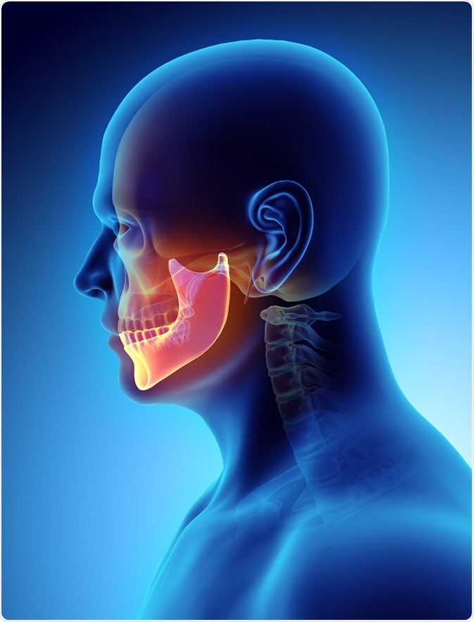
Jaw cysts are sack-like pouches that fill with fluid and form within the tissues of the jaw. These growths are not just limited to the jaws, because they can form anywhere within or on the body. Jaw cysts are generally benign in nature and non-cancerous growths, but may present with malignant degeneration very rarely. Cystic jaw lesions tend to grow very slowly and in many patients, they are asymptomatic (i.e. they do not cause any noticeable symptoms).

Credit: yodiyim/Shutterstock.com
Due to this nature, they are mostly found incidentally when radiographic imaging is conducted for another unrelated head and/or neck pathology. However, if the cysts become infected then they may evolve into painful entities.
Types of jaw cysts
There has been several classification systems used to categorize cystic lesions of the jaw. One classification by the World Health Organization groups these lesions into odontogenic and non-odontogenic cysts.
The majority of jaw cysts are termed odontogenic cysts, because they develop from dental tissue epithelium and as a result are delineated from non-odontogenic cysts (i.e. those with epithelium that is non-dental in origin). Odontogenic cysts may be further subcategorized based on their pathogenesis into inflammatory or developmental cysts.
Odontogenic cysts
Radicular cysts are inflammatory odontogenic cysts that arise from infection or trauma and are also known as periapical cysts. They are the commonest jaw-occurring cysts and present mainly between the third and fifth decades of life.
These cysts arise as the ultimate result in the pathway of tooth inflammation and necrosis of the pulp. On radiographic imaging, these lesions measure less than one centimeter in diameter and are lucent in the periapical area with a fairly round shape.
Eruption, follicular, gingival and glandular odontogenic cysts are all examples of developmental odontogenic cysts. Eruption cysts tend to disappear spontaneously and originate on the mucosa of teeth just before they erupt.
Follicular cysts are also known as dentigerous cysts, which form by fluid accumulation between the crowns of teeth that have not yet erupted and reduced enamel epithelium. They are the second commonest cystic lesion of the jaw and, present most frequently between the second and fourth decades of life, thus being almost exclusive to secondary dentition.
Gingival cysts occur frequently in newborns on the alveolar mucosa as vestiges of the dental lamina. The vast majority of these cysts occur in the mandible and they disappear spontaneously by rupturing into the oral cavity.
Gingival cysts may also occur in adults, most frequently within the fifth and sixth decades of life and arise from the rest cells of the dental lamina. Glandular odontogenic cysts are very rare, mostly slow-growing intraosseous lesions that generally tend to occur on average around the fifth decade of life and in the anterior mandible.
Non-odontogenic cysts
There are many examples of non-odontogenic cysts, which are the remnants of epithelium and the embryonic ducts that are left behind after embryogenesis. These cysts tend to be found deep in the areas where the epithelial ridges and walls of embryogenesis, as well the fissures and clefts, once were.
Non-odontogenic cysts may arise without or with inflammatory stimuli and two examples of these are nasolabial and nasopalatine duct cysts. The former cysts arise from the vestiges of Hochstetter’s epithelial wall, which plays a role in the formation of our noses, whereas, the latter arise from the entrapped leftovers of the nasopalatine duct during palatal plate fusion.
References:
- http://pubs.rsna.org/doi/full/10.1148/radiographics.19.5.g99se021107
- https://www.ncbi.nlm.nih.gov/pmc/articles/PMC3612207/
- https://radiopaedia.org/articles/dentigerous-cyst
- https://www.ncbi.nlm.nih.gov/pmc/articles/PMC4065455/
- https://radiopaedia.org/articles/incisive-canal-cyst
- https://radiopaedia.org/articles/nasolabial-cyst-2
Further Reading
Last Updated: Mar 21, 2019