Obstructive sleep apnea (OSA) is a sleeping disorder in which during sleep, the affected individual repeatedly stops and starts breathing.
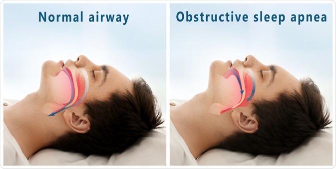
Image Credit: Africa Studio / Shutterstock.com
OSA and hypoxia
OSA is caused by the sleep-induced relaxation of throat muscles that support the soft palate, uvula, tonsils, and tongue, causing complete (apnea) or partial (hypopnea) blockage of the upper airway. This blockage prevents air from flowing into the lungs and results in insufficient oxygen levels in the blood and organs—a condition known as hypoxia.
Hypoxia triggers a breathing reflex in the brain that briefly awakens the person from sleep in order to resume normal air flow. Pauses in breathing typically last from 10 to 30 seconds but can continue for up to a minute. In severe cases, up to 50 or more breathing pauses can occur in a one-hour period. People who suffer from OSA frequently snore loudly and sometimes make snorting or choking sounds.
Risk factors for OSA include age, smoking, alcohol consumption, hypertension, and chronic nasal congestion. The causes of OSA can be exacerbated by sleeping position, with back sleepers suffering from more frequent episodes.
Risk factors
Although OSA can occur in people of all ages, it is typically seen in middle-aged and older adults. In particular, there is an increased risk for developing OSA in males, postmenopausal women, those of certain ethnic backgrounds, as well asthose with family history.
It is estimated that over 50% of all OSA sufferers are overweight; therefore, obesity is a significant risk factor for this condition. OSA is particularly common in those with a thick neck, which is defined as a circumference of greater than 17 inches for men and 16 inches for women, where fat deposits can inhibit airflow. One study has shown that even modest weight loss can lessen the number of apnea or hypopnea episodes per hour and lead to improved sleep. The worldwide prevalence of OSA is expected to grow as the number of people with obesity continues to rise.
Understanding Obstructive Sleep Apnea | Access Health
OSA occurs in people of all ages with craniofacial abnormalities or who possess anatomical features that make them prone to airway blockage. This includes people with large tongues, a small lower jaw, or Down syndrome.In addition, certain medical conditions such as a deviated septum, enlarged tonsils or adenoids, or chronic nasal congestion can lead to airway obstruction and interrupted sleep patterns.
OSA is also found in children, as between 1% and 10% of children are estimated to experience OSA. These occurrences typically develop during ages 3-6 and are caused by enlarged tonsils and adenoids. The surgical removal of the tonsils and adenoids is therefore the treatment of choice for this condition in children. As with adults, some children also experience OSA due to craniofacial abnormalities or obesity. In these cases, OSA symptoms can be alleviated with weight loss, continuous positive airway pressure (CPAP), and surgery, similar to adults.
Association with cardiovascular disease
OSA is a potentially serious medical condition because fluctuating blood oxygen levels put pressure on the heart and vasculature. Therefore, OSA sufferers typically exhibit increased risked factors for cardiovascular disease such as hypertension, insulin resistance, dyslipidemia, and inflammation. Several studies have confirmed the link between OSA and cardiovascular disease, concluding that people with OSA are more likely to suffer from hypertension, coronary artery disease, stroke, and heart failure.
References
Further Reading
Last Updated: Mar 17, 2021
 Tirzepatide vs. semaglutide: Study compares cost and health outcomes in obesity