Otitis media is a medical condition that is translated from Latin to mean “inflammation of the middle ear.” This infection occurs in the area between the eardrum and the outer ear and can be very painful.
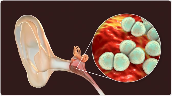
Image Credit: Kateryna Kon / Shutterstock.com
Epidemiology
Otitis media is a common condition, as approximately 11% of the worldwide population experiences at least one episode of acute otitis media each year. Half of all cases occur in children under the age of five, with males being more affected by otitis media than females.
A possible complication of the condition is chronic suppurative otitis media. It is estimated that nearly 1 in 20 people affected by an acute episode of otitis media will develop the chronic condition, which can last for several weeks to months.
The vast majority (80%) of children will experience otitis media with effusion at some point in their lives before reaching the age of ten years. In fact, otitis media is the most common condition that needs medical care in the United States for children under the age of five.
Risk factors
There are some factors that have been linked to a higher risk of otitis media. This includes children who are exposed to polluted environments, particularly if the parents smoke at home. Additionally, using a pacifier rather than breastfeeding and attending daycare from an earlier age with many attendees can have put children at an increased risk of acquiring otitis media.
Therefore, to help decrease the risk of otitis media, it is recommended that parents limit smoking in the home environment. Mothers should also be encouraged to breastfeed for as long as possible and, if daycare is required, centers with fewer attendees should be chosen when possible.
Symptoms
Ear pain is the characteristic symptom of otitis media, which is sometimes also described as a feeling of fullness in the ear. This may also cause people to become irritable, easily fatigued, and have a decreased appetite.

Image Credit: Sokolova Maryna / Shutterstock.com
When the condition is particularly severe or inadequate treatment has been administered, it is possible for the tympanic membrane to rupture. Whilst this is often accompanied by a feeling of relief as the pressure inside the ear is released, this can lead to excessive draining of the ear and increases the individual's susceptibility to severe infection inside the ear.
Additionally, it is possible for hearing loss to occur, which may be temporary or permanent, depending on the circumstances of the event.
Treatment
The majority of cases of acute otitis media tend to resolve on their own within a few days. For this reason, a simple analgesic for pain relief is often a sufficient treatment for many cases of acute otitis media. This helps to reduce pain and irritability in children while the infection passes of its own accord.
However, for people with particularly severe pain and fever, or for those less than 24 months old, treatment with antibiotics should be initiated.
Amoxicillin is the treatment of choice for most patients; however, amoxicillin-clavulanate is a suitable alternative if there is suspected resistance to amoxicillin or if it has already been used in the previous 30 days.
An improvement of symptoms should be evident within 2 to 3 days. If the condition does not appear to be improving, it is recommended to add or change the treatment regime, as the current therapy is unlikely to be offering a large benefit.
References
Further Reading
Last Updated: Dec 21, 2022
 World’s largest eye imaging and data resource expands across the UK