Bile duct cancer is identified, diagnosed, and staged with the help of tests that analyze bile ducts and neighboring organs. To find, diagnose, and confirm bile duct cancer, several different tests and procedures are generally performed.
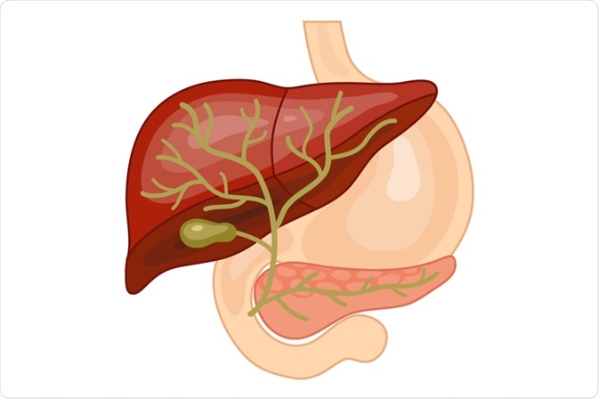
Image Credit: marina_ua / Shutterstock.com
Diagnosis
A general physical examination of the body is carried out to examine the patient's health signs, including signs of ailment, such as any abnormal condition or formation of lumps. This physical exam will also include exploring the lifestyle and medical history of the patient.
Upon the identification of something suspicious, several laboratory tests will be ordered. These tests often involve examining blood, tissue, urine, or additional components present in the body. With the help of these tests, the disease can be diagnosed quickly to allow for healthcare providers to begin determining the appropriate treatment approach for the patient.
In addition to some of the traditional laboratory tests that will be ordered, a CA 19-9 tumor marker test and carcinoembryonic antigen (CEA) test may also be prescribed. In this procedure, the volume of certain components produced by tumor cells, tissues, and other organs in the body is measured in a sample of tissue, urine, or blood.
The presence of increased levels of certain components in the body that can relate to specific cancer types which, taken together, are referred to as tumor markers. In bile duct cancer, for example, abnormal levels of CA 19-9 and CEA, the latter of which is also known as a carcinoembryonic antigen, are often present.
Imaging
Ultrasonography is often used to confirm a diagnosis of bile duct cancer. In this method, high energy sound waves known as ultrasounds produce echoes as they deflect off interior tissues or organs like the abdomen. A sonogram is the image of body tissues that are generated from the echoes, the images of which can be printed for later use.
A sequence of accurate images of interior body sections, especially the abdomen, is captured at different angles during a computed tomography (CT) scan. The images are generated by a computer that is connected to an X-ray machine. For more detailed views of tissues and organs, contrast agents may be utilized.
When obtaining a magnetic resonance imaging (MRI) scan, a sequence of images of the internal body parts is generated through the use of radio waves, a magnet, and a computer.
Magnetic resonance cholangiopancreatography (MRCP) is another technique that can be used during the diagnosis of bile duct cancer. To this end, MRCP obtains a sequence of images of interior body parts such as the bile duct, pancreas, liver, gall bladder, and pancreatic duct through the use of radio waves, a magnet, and a computer.
Understanding Liver and Bile Duct Cancer
Noninvasive biopsies
Following the completion of the aforementioned diagnostic procedures, a needle biopsy may be indicated to further understand the types of cells present in bile duct cancer. In this method, without making a surgical cut initially, a fine hollow needle is infused into the tumor through the skin.
A local anesthetic is initially used to desensitize the skin. Generally, the needle is directed into the spot by using a CT scan or ultrasound. When the picture displays that the needle is pointed towards the tumor, a specimen is drawn into the needle and is then sent to pathology for further microscopic evaluation.
A fine-needle aspiration (FNA) is another biopsy technique that used in many cases. During an FNA, surgeons will utilize a very thin needle connected to a syringe to withdraw cell samples. It is rare that the FNA will not provide sufficient cells for an accurate diagnosis. If this is the case, however, more samples can be collected by making use of a large needle by performing a core needle biopsy.
Invasive biopsies
Several different surgical procedures may also be indicated in bile duct cancer cases. These include laparoscopy, endoscopic retrograde cholangiopancreatography (ERCP), and percutaneous transhepatic cholangiography (PTC).
Laparoscopy
A laparoscopy is a surgical procedure that is used to examine the interior portion of the abdomen, such as the liver and bile duct, to further investigate the area for any signs and symptoms of cancer.
During this procedure, tiny cuts are made in the abdominal wall and a thin tube fitted with light known as a laparoscope is inserted through one of the incisions. To confirm the presence of cancer, samples of tissues are collected by inserting the equipment either through the incisions.
ERCP
Bile is carried from the liver to the gallbladder and then from the gallbladder to the small intestine. The ERCP method is used to obtain X-ray images of these ducts.
Typically, these ducts tend to be confined and the flow of bile is restricted or slowed down, which leads to jaundice. By using an endoscope, the camera is able to reach the small intestine by passing through the mouth and abdomen.
PTC
PTC is a method used to X-ray the bile ducts and the liver. During this procedure, a thin needle is infused into the liver through the skin beneath the ribs. X-ray images are then taken by infusing the dye into the bile ducts or the liver. To check for signs of cancer, tissue samples are collected.
The stent is a thin soft tube that may remain in the liver to drain out bile into the small intestine. In the event that the bile duct is obstructed, a collection bag will be placed outside of the body to allow for the drainage of the bile from the bile duct. This method is often used if surgery is not a suitable option for the patient.
References
Further Reading
Last Updated: Dec 21, 2022