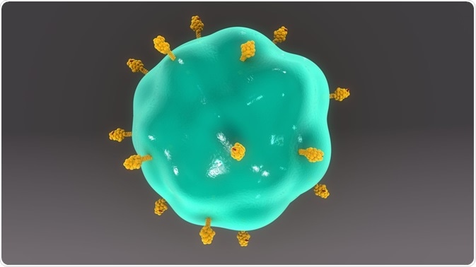
Flow cytometry can be used to analyze different cell types. This technique can be applied to immunology to ensure that, prior to a transplant procedure, interactions between donor and recipient immune components do not cause an adverse immune reaction.
 sciencepics | Shutterstock
The role of HLA proteins
The human leukocyte antigen (HLA) proteins are crucial for the body’s immune defense against potentially harmful foreign substances. Class I and II HLA (HLA I and II) proteins are the ones involved with the immune response and transplantations. HLA I proteins are expressed on the surface of all cells. Conversely, HLA II proteins are expressed on the surface of antigen presenting cells (APCs) of the immune system, such as Dendritic cells and B-Cells.
Antigens and proteins are broken down into peptides, and these peptides form a complex with HLA proteins. The HLA-peptide complex subsequently interacts with effector T-cells causing intracellular signals in both cells, which determines if a specific immune response occurs. Effector T cells differentiate between ‘self’ and ‘non-self’ proteins - therefore, if the peptides presented are recognized as ‘non-self’, an immune response will commence.
HLA proteins are highly varied, as three genes contribute to the formation of HLA I proteins, while six genes contribute to the formation of HLA II proteins. Given that there are two distinct alleles for each gene, the possible combinations are numerous.
Flow cytometry
Flow cytometry represents an analysis technique that can be used to study both the physical and chemical properties of cells and/or particles. During flow cytometry, the sample is suspended in a fluid and injected into the flow cytometer. Usually, one cell at a time is passed through a laser beam for analysis purposes. The scattering of light caused by this gives information on the characteristics of the cells of the sample.
The cells are fluorescently labeled before being passed through the cytometer. The labels contain antibodies which are attached to fluorochromes. Separate labels can be used for different cells within a sample, which allows for a heterogeneous population to be analyzed. Isotopes can also be attached to the antibodies, which is usually seen during mass cytometry.
Flow cytometry cross-matching
Flow cytometry cross-matching (FCXM) involves mixing donor lymphocytes, the recipient’s immune serum, and fluorescent labeled antibodies into a sample. The antibodies used are specific to the donor HLA and various T-cell and B-cell specific markers (e.g. CD3, 5, and 8 for T-cells, and CD19, 20, and 21 for B-cells).
The sample is the ran through a cytometer, so the lymphocytes can interact with the antibodies in the recipient’s serum. If there are donor-specific HLA antibodies in the serum, they will bind to the donor lymphocytes, which allows the fluorescently labeled antibodies to bind, giving in turn a positive cross-match.
The benefits of flow cytometry cross-matching before transplantation
A problem with allograft transplants is that organ rejection can occur, which is seen as a result of the recipient’s antibodies against the donor HLA. This will elicit an immune response which can subsequently cause rejection.
Since a positive FCXM is associated with an increased chance for a transplant recipient to reject the prospective transplant, FCXM is important, as it allows for donor antigens and host antibodies to be checked prior to a transplant to avoid a host immune response. This reduces the chances of acute or chronic allograft rejection.
Complications with flow cytometry cross-matching
During flow cytometry, the antibodies that are used can bind non-specifically and give unreliable results. This can be caused by protein-protein interactions, glycolipid interactions, electrostatic interactions, and binding to Fc receptors.
The use of a pre-digestion agent can reduce the chances of non-specific binding, which increases the sensitivity and reliability of the analysis. Finally, combining other analytic techniques (such as sera absorption) with FXCM can also reduce the rate of false-negative results.
Further Reading
Last Updated: Jun 25, 2019