Proteins, also called polypeptides, are the polymers of amino acids. There are a total of twenty amino acids called monomers that exist naturally in proteins. Proteins are found in abundance and are differentiated from each other according to the number, type, and arrangement of amino acids in series, which comprise the mainstay of polypeptides.
Credit: PolakPhoto/Shutterstock.com
For different reasons, proteins are the main components of food, as well as an important source for producing energy. Proteins are generally present in many natural food types such as fish or meat. Many of the proteins in food are enzymes that are able to improve a certain number of biochemical reactions. The presence of amino acids in proteins is very essential for human health.
Methods involved in analyzing protein
Different proteins have different molecular structures, physiochemical properties, and nutritional attributes. Three major steps are involved in protein analysis.
1. Protein separation
The following are some of the common methods for separating proteins:
- SDS (sodium dodecyl sulfate)-PAGE: Protein separation by this method is based on the molecular weight, as opposed to fold or charge. Factors other than the molecular weight, however, may also affect the movement of proteins on SDS gel, which was analyzed in a course group. This technique is broadly used in genetics, biochemistry, molecular biology, and forensics.
- Isoelectric focusing: Separation of protein by this method is dependent upon balance between the negative charged (acidic) and positive charged (basic) constituent amino acids, which defines a property called protein isoelectric (PI) point. The net charge of the proteins in pH becomes accurately zero. Proteins that vary by as small as a single charged residue can be isolated by this method.
On the other hand, however, proteins of different sizes with a similar PI value can be placed at the same place. Often, two chromatic methods are used for the separation of proteins or even for peptide separation.
- High-performance liquid chromatography (HPLC) methods are used for separating and purifying peptides or proteins on the basis of charge, size, or on the whole hydrophobicity.
- Thin-layer chromatography (TLC) has also been used for separating peptides on the basis of their similar properties.
- Gel Electrophoresis through two-dimensions (2D): This is a powerful technique generally used for analyzing complex samples, but it may not be used for a few or specific proteins, in the interest of characterizing all varieties of protein samples. In this method, first the protein is passed in a narrow path of isoelectric targeting gel. Based on the isoelectric point the protein can be separated.
Then the isoelectric targeting gel is situated over the SDS-PAGE gel and it is run in the perpendicular direction. Proteins run as spots and their position is based on their size and charged properties. This provides a powerful separation of proteins using either of these two gels only, which are isoelectric focusing or SDS-PAGE.
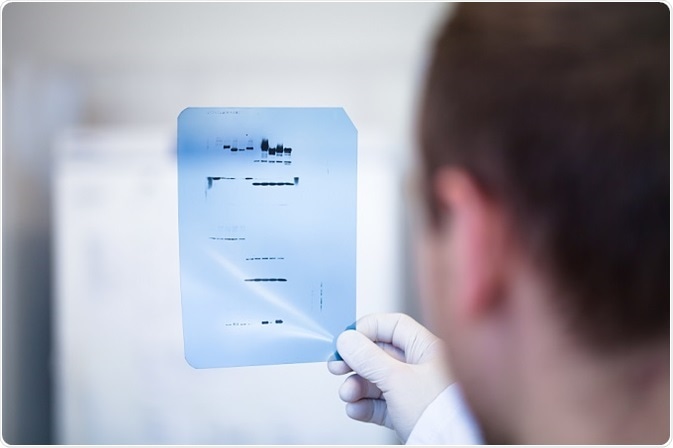
2. Western blotting
Immunoblotting is considered the most general version of western blotting and is used in combination with chromatographic methods. This method is used for identifying specific proteins within a given piece of tissue extract or homogenate. The protein sample is first electrophoresed by using SDS page for the separation of proteins on the basis of molecular weight.
Following this, the wet gel is positioned against the nitrocellulose sheet and kept in a special kind of electrophoretic chamber. Thereafter, an electric field is passed into the gel that causes the protein to move out of the gel and also onto the sheet of nitrocellulose in which it becomes adsorbed tightly. Afterward, nitrocellulose sheet with this tightly bounded protein can be “probed” or “blotted” with antibodies, specifically for targeting the protein.
3. Protein identification
If the protein spot or band was found on a gel, the next step is to determine their molecular identity. The two methods commonly available for this method are as follows.
- Edman degradation: This method was developed by Pehr Edman. He used reagents called isothiocyanates to act and react with the N-terminus of the peptide or protein. Under certain suitable conditions, a solitary “round” of Edman degradation can split the amino terminus residue to produce amino acid derivative with the addition of a new free N-terminus. Eventually, the amino terminus is separated from the protein, without interrupting the bonding of proteins between the other residual amino acids.
- Mass spectrometry: This is the most apt method for identifying the exact protein molecular weight. Because this method is very sensitive and amenable to the quick processing of several samples, this technique has become popular for identifying the different kinds of proteins that are present in a complex sample. Hence, mass spectrometry may be used for determination of the sequence of the proteins and thus the protein identity in a way conceptually like the above described, based on an Edman degradation.
Reviewed by Afsaneh Khetrapal BSc (Hons)
Last Updated: Jul 19, 2023
 WuXi Biologics and Hanchorbio enter strategic partnership to advance next-generation bi- and multi-functional fusion protein pipeline