Chromatography is the separation of compounds from a mixture, and this is dependent on the properties of the compound. These properties include the size and charge of the compounds, and also how they interact with the mobile and solid phases.

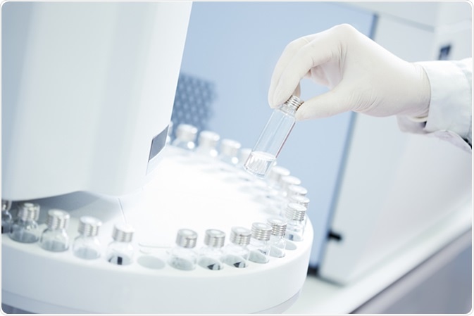
Technician loading sample vials in autosampler rack. Image Credit: Stokkete / Shutterstock
Affinity and Pseudo-Affinity Chromatography
Affinity chromatography is where a specific ligand is added to the solid phase, which captures the compound of interest like a specific protein. The specific nature of this interaction means that the protein of interest can be separated from a mixture of proteins. The protein of interest can then be subsequently released from the ligand by changing the conditions of the chromatography.
In similar fashion, pseudo-affinity chromatography utilizes dyes as ligands in order to target proteins. The dyes used mimic the ligands, but they do not display high specificity. Therefore, these dyes are able to capture a variety of proteins.
How Does Pseudo-Affinity Chromatography Work? – An Example
A study by Tulsani and co. looked at using pseudo-affinity chromatography to separate three enzymes from rabbit muscles: lactate dehydrogenase, pyruvate kinase and aldolase.
Here, cross-linked guar and cross-linked pectin were used to immobilize various triazine dyes. To immobilize the dye, first the cross-linked guar and cross-linked pectin were washed in deionized water, and then suspended in fresh deionized water. The dye was then added to the gel, and this was stirred for about 5 mins. Salt was added to this mix, and then the mix was stirred for a further 30 mins. Sodium hydroxide was then added to the mix, and this was stirred for 16 – 18 hours. Finally, this mixture was washed in deionized water.
The gel, now with the dye bound, was placed into 2ml columns. Once phosphate buffer had passed through the column, the crude protein sample was loaded onto the top of the column. Once the sample had passed through, the column was washed with the buffer to remove the proteins that were unbound. This was verified by measuring the absorbance of the buffer leaving the column at 280nm using a spectrophotometer. Once clean, the bound protein was released by using the same buffer, but with added salt, NAD+ or sodium pyruvate.
The process managed to separate lactate dehydrogenase, pyruvate kinase and aldolase. It was found that dyes 1014 and 1015 retained lactate dehydrogenase, and 1015 bound to cross-linked pectin also retained pyruvate kinase. However, aldolase was not retained by any of the 10 dyes tested in this study.
Removing Proteins from Plasma
There are proteins present in human plasma, which is the liquid component of blood. This is a highly complex mixture, of which 75% are human serum albumin and immunoglobulin G (IgG), a type of antibody. This means that those proteins are very easily detected, but also that they could prevent other proteins from being detected. This could be problematic, as diagnostic markers for certain diseases could be those low-abundance proteins. Therefore, the removal of serum albumin and IgG is a crucial step in preparing serum for diagnostic purposes.
A study by Urbas and co. looked at optimizing the methods used to remove serum albumin and IgG from plasma. Here, they not only used pseudo-affinity chromatography to selectively remove serum albumin, but combined this with affinity chromatography so that both serum albumin and IgG could be removed at the same time. While this was found to be efficient, the Mimetic Blue SA A6XL dye used in the pseudo-affinity chromatography to remove serum albumin was not highly specific. Therefore, the ability of this system to remove serum albumin depended on the amount of plasma loaded onto the column. If more plasma was loaded onto the column, the increased amount of serum albumin was able to displace the other proteins from the dye.
Further Reading
Last Updated: Aug 12, 2022