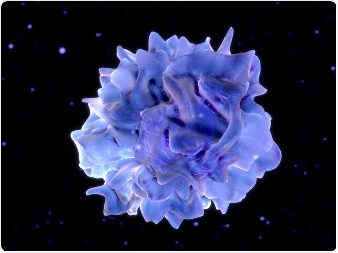
Flow cytometry is a technique in which multiple parameters of a single cell or molecule are measured as it passes through a detector in a liquid suspension. It is based on the way light scatters when it passes a laser beam.

Juan Gaertner | Shutterstock
Various studies have looked at the possibility of studying cell receptors using flow cytometry. These types of studies can involve receptor-specific antibodies, fluorescent receptor ligands or a genetic tag.
Using fluorescence microscopy to study the dopamine receptor
Study by McKenna and co. used flow cytometry to see if dopamine receptors could be detected on white blood cells. Dopamine receptors fall into D1-like (D1, D5) and D2-like (D2, D3, D4), and all except D1 was able to be detected by flow cytometry.
Of all the white blood cells tested, T-lymphocytes and monocytes were found to express low levels of dopamine receptors, neutrophils and eosinophils had moderate expression and B-lymphocytes and NK cells expressed high levels of dopamine receptors.
Identifying naïve T cells
Another study showed that the loss of naïve CD8+ and CD4+ T-cells were hallmarks of HIV progression. The authors then decided to take this further and develop a flow cytometry-based method for differentiating naïve T-cells, which was used to analyze the diversity of T-cell receptors and detect the loss of CD8 and/or CD4.
What about the function of receptors?
Chemokines are “inflammatory” or “homeostatic”; inflammatory chemokines recruit leukocytes to the site of infection or damage, while homeostatic chemokines regulate the movement of leukocytes in specific tissue. Various studies have used flow cytometry to study the function of chemokine receptors
Chemokine receptor trafficking
One example is the study of how chemokine receptors are “trafficked”. In this instance, trafficking refers to the internalization, lysosomal degradation/deactivation and subsequent recycling of the receptor to the cell surface.
A process referred to as “antibody feeding” can be adapted for use by flow cytometry to study chemokine receptor trafficking; here, a primary antibody specific to the receptor of interest is added to cells, then exposed to the ligand (or not in the case of controls).
A secondary antibody with a fluorescent molecule is then added, and the fluorescence on the cell surface can be measured by flow cytometry. An equation can then be used to determine the percentage of receptors which were on the cell surface.
For this, it is important that the primary antibody is not only specific, but also that it does not bind to the ligand binding site or the activation site. This ensures that the ligand or receptor activation does not interfere with antibody binding, and that the only loss of antibody binding is through internalization of the receptor.
Receptor signaling
Chemokines binding to their receptors cause the activation of signal transduction pathways, and one change this causes in cells is the amount of calcium in the cytoplasm. Activation of chemokine receptors by binding of the chemokine leads to the release of calcium from its intracellular stores, which then causes calcium to enter the cell to replenish the stores.
Certain fluorescent dyes can change their characteristics when bound to calcium, so this combined with flow cytometry can be used to study how calcium is moving in the environment. In such studies, one or more of these dyes are added to cells, which are then activated by the addition of the chemokine. A change in fluorescence can then be detected by flow cytometry.
Further Reading
Last Updated: Dec 18, 2018