LASER (Light Amplification by the Stimulated Emission of Radiation) has a wide range of uses in dentistry.
Lasers in hard dental tissue are primarily used for prevention of cavities, bleaching, removal of an unwanted filling material, growth modulation, treating dentinal hypersensitivity, and for diagnosing dental problems.
Over the past two decades, the application of lasers has shown its effectiveness in terms of its efficiency, precision, and relief from pain in complex dental treatments.

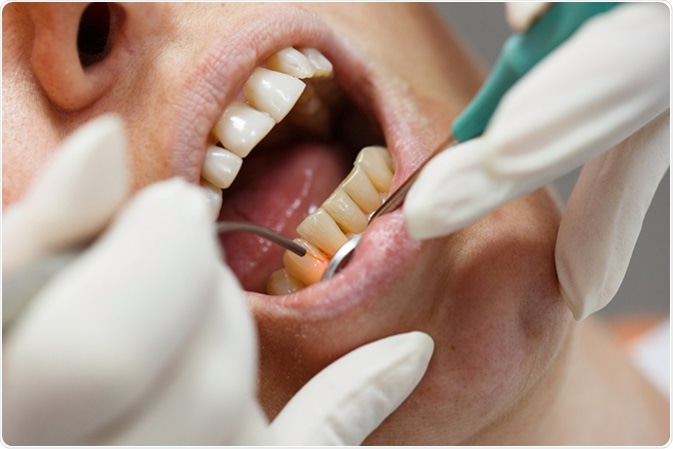
Dental laser used on a patient on soft and hard tissue. Image Credit: zlikovec / Shutterstock
Types of Lasers Used in Dentistry
There are different ways by which lasers are used in dentistry. Lasers are categorized by various methods, however broadly, they are classified according to:
- Lasing Medium: Gas or Solid
- Tissue applicability
- Hard or Soft tissue lasers
- Range of Wavelength
Varieties of Lasers
Carbon Dioxide (CO2) Laser
The CO2 laser wavelength has a high attraction for water, which allows fast soft tissue elimination with minimal penetration. The CO2 laser has high tissue absorption. However, it is not preferred primarily because of its large size, hard tissue destruction, and expensive cost.
Erbium Laser
Erbium Lasers have two subtypes depending on the wavelength: Er, Cr: YSGG (yttrium scandium gallium garnet) lasers and Er: YAG (yttrium aluminum garnet) lasers. By far, these lasers have shown the most promising results for application in issues related to dental hard tissues.
e.Max Veneer Removal with Hard Tissue Laser
Neodymium Yttrium Aluminum Garnet Laser (Nd: YAG)
Nd: YAG Lasers are most effectively taken in by the pigmented tissue, hence they are primarily used for soft tissue surgeries.
Laser Periodontal Therapy - WPT™ Animation
Diode Laser
Diode laser consists of aluminum, arsenide, gallium, and rarely indium, which collectively creates laser wavelengths.
The Principal of Laser Functioning
The laser is a monochromatic light which basically consists of three parts:
An Energy Source
The energy source pushes energy (e.g. electrical current) in the system through a device (e.g. electrical coil).
An Active Lasing Medium
The energy generated from the source is then pushed through an active medium which is placed between optical resonators. This leads to the emission of photons. Laser properties such as wavelength are dependent on the type of medium (e.g. gas, crystal, semiconductor) that is being used.
A Resonator
The emitted photons are then amplified as they are reflected back and forth between resonators (e.g. two or three mirrors) through the active lasing medium before their release to the target area.
In dental surgery, the laser is guided to the target area or tissue area either through a fiberoptic cable, hollow waveguide, or an articulated arm.
Once the tissue absorbs the laser its temperature increases, which leads to photochemical effects. But, this effect is dependent on the water content of the target tissue area. When the temperature rises over 100 degrees water from the target tissue start to vaporize. This process is termed as ablation.
Applications of Lasers in Dental Hard Tissue
Dental Restorations and Caries Prevention
Argon Laser releases high intensity visible blue light, which has the capacity to start the photopolymerization process of light-cured dental fillings. Light cure dental fillings are tooth-colored fillings and with the help of high-intensity blue light, they adhere to the tooth surface. Aside from that, argon laser has an excellent ability to change the surface properties of dentin and enamel, which protects the tooth against cavities.
Laser Fluorescence
At times, orthodontic treatment leaves certain white spots post-removal of the fixed appliances. These white spots occur as a result of demineralization. Application of lasers has shown a reversal of demineralized spots.
Removal of Restoration and Cavity Preparation
Er: YAG, have been used for caries removal from enamel and dentin portions of the tooth through the process of ablation without damaging the pulp of the tooth. Er: YAG can also help in cavity preparation for filling the tooth with restorative material.
Also, in case if an unnecessary tooth filling requires removal, Er: YAG can then be used for the same.
Etching
Etching is a technique in which a smooth enamel surface is converted into an irregular and rough surface.
Acid etching is a conventional method used during tooth restoration procedure. But, Er: YAG lasers have emerged to be a substitute for the acid etching technique.
Dentinal Hypersensitivity
Application of Er: YAG has shown good results in treating dentinal hypersensitivity. Er: YAG is more effective and long lasting as compared to conventional treatments when treating the cervical portion of the tooth which has hypersensitivity.
Diagnosing Dental Problems
Lasers have come up with diagnosing various dental diseases like caries detection by optical changes and profiling of tooth surface and dental restorations.
Growth Modulation
Laser scanner produces high-quality 3D images which give a detailed view of craniofacial structures during different phases of growth and development.
These non- destructive 3D images of oral dental structures are also helpful in analyzing clinical outcomes in dental treatments.
Advantages and Disadvantages of Lasers
Application of Lasers is emerging in the field of dentistry. Use of lasers reduces the pain, minimizes the chances of bacterial infections and ensures quick healing during hard tissue treatments.
Also, patients may experience less bleeding, there are no sutures, and tissues regenerate faster compared to conventional methods.
Patients also experience less anxiety during most of the hard tissue dental procedures. However, lasers have certain drawbacks such as their limited scope to reach all areas requiring dental treatments, tissue damage, and high cost.
Further Reading
Last Updated: Mar 13, 2023