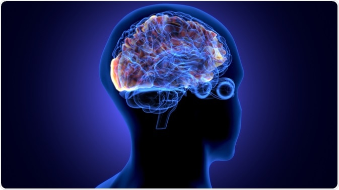
Menopause is a state of reproductive senescence in the female. It begins in middle life, involving both neurological and endocrine aging. It passes through different stages and is caused by the decline in female sex hormones.
 Image Credit: Life science/Shutterstock.com
Background
Menopause-related drops in function may be the result of natural aging of the endocrine axis or caused by the removal of the ovaries or medical treatment that prevents ovarian endocrine function.
However, menopause is also a neurological change, as shown by many hallmark symptoms of menopause, especially forgetfulness, sleep disturbances, altered mood, and hot flashes. Ovarian and brain health are therefore inextricably linked in women.
As estrogen levels fall, changes occur in the morphology, number, and interactions between nerve cells, their glucose metabolism, and gene expression. In female animal models, low estrogen has been linked to the accumulation of the abnormal protein amyloid-beta (Aβ), which is notorious for forming plaques within brain tissue, in people with Alzheimer’s disease (AD), though also sometimes in normal people.
Brain-endocrine axis
Depending on the stage of menopause (pre-menopause, peri-menopause, and post-menopause), the brain structure and neural connectivity, as well as energy metabolism, undergo marked changes. The brain areas involved in higher cognitive functions were most affected at all ages.
This effect was observed independent of other cardiovascular or dementia risk factors such as ApoE-4 levels, hysterectomy, or the use of hormone replacement.
In post-menopausal women, the earliest change in the brain appears to be a fall in the amount of glucose used by the brain, indicating reduced brain activity. This is due to falling estrogen levels, this hormone being vital for brain glucose metabolism.
Neuroscientist Lisa Mosconi, the author of a study on this aspect of menopause, explains: “When estrogen doesn't activate the hypothalamus correctly, the brain cannot regulate body temperature correctly. So those hot flashes that women get - that's the hypothalamus. Then there's the brainstem in charge of sleep and wake. When estrogen doesn't activate the brainstem correctly, we have trouble sleeping. Or it's the amygdala, the emotional center of the brain close to the hippocampus, the memory center of the brain. When estrogen's levels ebb in these regions, we start getting mood swings perhaps and forget things.”
Gray matter volume in the brain declines as well. These parameters respond to hormone replacement, however, suggesting that the link between the central nervous system and endocrine axis remains active for a long time after the menopausal transition begins.
These markers of cognitive decline normalized after menopause, and the gray matter volume also recovered to baseline in the brain areas most concerned with menopausal endocrine aging.
Vasomotor symptoms and cognitive impairment
Cognitive decline is common during the transition into menopause, including symptoms such as forgetfulness and delayed verbal memory, reduced verbal processing speed, and impaired verbal learning. Earlier research shows that memory-related complaints are predicted by age, hot flashes, depression, feelings of stress, and the perceived level of health.
Healthy women demonstrated slight but consistent changes in verbal memory and learning, as well as processing speed. Reassuringly, performance returns to normal postmenopausally.
Vasomotor symptoms (VMS) are the symptom characteristic of the menopausal transition but may occur before, during, or after menopause. These symptoms include hot flashes and night sweats. Like cognitive symptoms, their occurrence does not mirror the falling levels of estradiol.
At present, it is thought that the hippocampus, parahippocampus, and multiple regions of the prefrontal cortex of the brain are involved in the origin of the cognitive decline associated with severe VMS.
VMS is associated with high blood pressure, high blood lipid levels, a tendency towards insulin resistance and diabetes risk, and sometimes a procoagulant profile. The risk of future stroke is also suggested to be higher in women with more severe objective VMS.
VMS is also associated with increased cortisol levels, at about 20 minutes following the event, which is known to be linked to memory impairment. These events may also often cause a 5% decrease in blood flow. Thus, VMS may be determinants of cognition at this midlife transition point.
VMS could thus be a risk factor for cardiovascular disease and cognitive impairment during this transition, as well as the key mediator between them.
Recovery after menopause
Compared to males of the same age, women undergoing the menopausal transition have significant alterations in brain biomarkers. The neurological changes occurring around this time cause symptoms that in turn trigger depression and anxiety, as well as AD, in a fraction of women.
The gradual onset of endocrine aging with spontaneous menopause may allow the brain to adjust to and compensate for the loss of estrogen and estrogen receptor activity. This resetting of the brain may explain why symptoms like hot flushes ease 2-7 years from their onset.
Neuroimaging confirms these findings. In the post-menopausal woman, gray matter volume returns to normal, especially in areas concerned with some types of memory and cognitive processing. In fact, in postmenopausal women, the gray matter volume was similar to that of males of the same age and increased over the next two years.
This volume also corresponded with rising memory scores in an area called the precuneus which shows structural changes during the menopausal transition. This is regulated by estrogen, and also changes during pregnancy, another unique female stage associated with neurologic and endocrine changes.
White matter volume falls during menopause and does not recover thereafter. However, compared to males, women undergoing or past menopause showed higher structural connectivity and myelination, which may indicate that the neural networks in these regions are more efficient following the onset of menopause.
Energy metabolism
Estrogen is key to glucose utilization in the brain. During the menopausal transition, the metabolism of glucose in the brain first dropped but then steadied at a new level, again suggesting adaptive changes. ATP levels rose after menopause, as did global measures of cognition. Mitochondrial recovery may explain how women are able to continue with intact cognitive abilities after menopause.
Hormone therapy and cognition
Does hormone therapy help preserve cognition? Dr. Mosconi says that hormones may help improve cognition. In late postmenopausal women, the risk of dementia is higher with combined estrogen-progesterone therapy, but unchanged when estrogen alone is used. In early post-menopause, however, hormones showed no effects on cognition.
She summarizes, “Overall, HT’s efficacy is thought to depend on timing of treatment initiation with respect to age at menopause, with benefits pertaining to early initiation, especially after induced menopause.”
Hormones may interact with high body weight to produce the opposite effect. Conversely, lifestyle and fitness may interact positively with estrogen, especially in the long term. Some research suggests that local estrogen synthesis within the brain occurs independently of estrogen in circulation after menopause, maintaining hippocampal function.
Conclusion
Menopause itself, therefore, does not herald a drop in cognitive ability, unlike the transitional period itself. In fact, women do better at cognitive tasks than men over their adult life, including during dementia! Instead, menopause may be considered “a dynamic neurological transition that reshapes the neural landscape of the female brain during midlife endocrine aging, [with] an adaptive process serving the transition into late life.”
Further studies will be necessary to understand how genetic factors and medical conditions affect cognitive function during menopause.
How menopause affects the brain | Lisa Mosconi
References
Further Reading
Last Updated: Sep 27, 2021