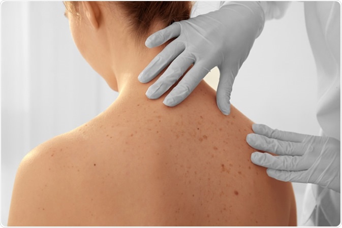
Mole mapping is a procedure used for the surveillance of skin for malignant melanoma. Skin is examined, and through the use of dermoscopy, lesions of concern are identified and evaluated.
Typically, mole mapping is done by means of photography or digital images of the whole body surface. This is kept as a record for future comparison to identify new lesions, or track changes in existing lesions.

Image Credit: Africa Studio / Shutterstock
There are automated systems available for mole mapping which are extremely accurate, but must be used in conjunction with a clinical examination by a doctor.
When caught early at a stage where it is confined to the epidermis, melanoma is considered to be completely curable. In fact, the survival rate for a melanoma less than 1 mm deep at five years is 94%. Comparatively, the five-year survival rate for patients with stage IV melanoma is less than 15 percent.
Early detection of melanoma can dramatically improve the rate of cures and survival in the disease; however, the early signs of melanoma are ambiguous, and the lesions can easily be mistaken for moles. For a person with a lot of moles, it may not be immediately obvious that a new lesion has appeared.
Studies have shown that mole mapping using digital photography can improve the early detection of melanoma. In one study, the chance of detecting melanoma increased by 17% when digital dermoscopy was employed. In another study, total body photography was found to improve the detection of new or subtle melanomas that did not fit the classical clinical profile of the disease.
Know your moles
Lesions of concern
Lesions of concern include those that resemble melanoma, basal cell carcinoma, or squamous cell carcinoma.
The ABCDE rule and the Glasgow 7-point checklist are used to identify potential melanoma lesions, although these guidelines may not catch early or atypical melanomas. As well, not every lesion identified through these instruments turns out to be a melanoma, and some of them are harmless.
Non-melanoma skin lesions, which include basal cell carcinoma and squamous cell carcinoma, are more common than melanoma. These lesions may appear crusty, ulcerated, or bleeding.
Candidates for mole mapping
People with more than 50 moles are candidates for mole mapping, as well as those with large or unusual moles. Mole mapping is also often used on the back, where it may be difficult for the patient to monitor them.
Patients with a family or personal history of melanoma may consider mole mapping, as well as those with fair skin who has been severely sunburned in the past. Any patient with a concern about specific moles or freckles may opt for mole mapping as a way of monitoring them for any changes in the future.
As compared to a skin self-examination, mole mapping produces a record that can be used to determine whether a lesion is new or has changed in any way. Growth or change in a mole can continue to be monitored by further mapping, or a physician may choose to intervene by removing the lesion.
Mole mapping may overlook lesions in hidden places on the body, like the genitals or the scalp. It can also give false negative or false positive results. Mole mapping can miss fast-growing melanomas, which attain a dangerous size before the next scheduled mapping session. Lastly, this monitoring procedure may not be as accurate with pink or scaly skin cancers, which do not show up as well in a photograph.
References
Further Reading
Last Updated: Mar 25, 2021