Skip to:
Mediastinitis is a term used to describe all inflammatory processes of the connective tissue of mediastinal structures and involves spaces between serous membranes known as pleura. Due to the vital nature of the aforementioned structures, this condition is associated with a significant degree of morbidity and mortality. Most cases of mediastinitis necessitate the admission and subsequent treatment in the intensive care unit (ICU).
A mediastinal infection has many possible causes. The three most common ones are deep sternal wound infection, perforation of the esophagus, and descending necrotizing mediastinitis. Spread via blood from a remote focal infection has been described but is rarely observed. In any case, most cases are associated with cardiac surgery, usually following coronary artery bypass graft.
In addition to these acute types of mediastinitis, a rare chronic form (sclerosing mediastinitis or chronic fibrosing mediastinitis) may be caused by a granulomatous infection. The condition is not bacterial in nature, but stems due to leakage of fungal cell constituents from lymphatic nodes into the mediastinum, resulting in an immunogenic reaction and ebullient fibrotic response.
The anatomy of the mediastinum
The mediastinum represents the region between the lungs, located behind the breastbone (sternum) and in front of the thoracic part of the spine (vertebral column). It is viewed as a central section of the thoracic cavity, extending to the diaphragm from the thoracic inlet. It can also be further divided into the anterior, middle, and posterior mediastinum.
The anterior mediastinum includes the thymus gland, adipose tissue, lymphatic nodes, and internal mammary vessels. The middle mediastinum is bounded by pericardium and includes the heart and big vessels, the trachea and bronchi, lymphatic nodes, as well as essential nerves. Finally, the posterior mediastinum contains the esophagus.

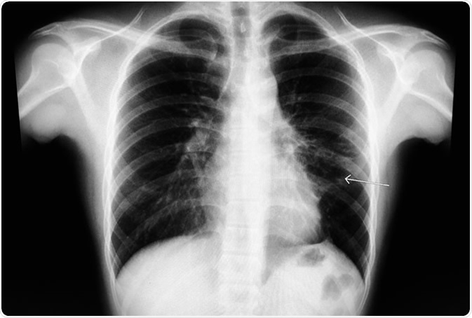
Chest X-ray known case S/P left anterior mediastinum mass. The study shows recticulo-fibrosis at medial perihilar LUL, and small tenting LLL basal fibrosis. Cardiac contour and rib cages are intact. Image Credit: Good Image Studio
Epidemiology of mediastinitis
The incidence of the condition after surgery ranges from 0.25% up to 5%, but it may differ according to the institution, as well as the type of procedure pursued on a patient. A majority of large centers with ample expertise and experience report rates that are lower than 2%. Reported mortality rates from mediastinitis vary significantly.
The condition is known to occur at any age; however, it is most commonly seen in older patients following thoracic or cardiac surgery. The most significant comorbidities that may predispose patients for the development of acute mediastinitis are obesity, diabetes, smoking, obstructive lung disease, peripheral arterial disease, but also prior heart or thoracic surgeries.
Mediastinitis in children usually arises as a complication of either thoracic or cardiovascular surgery but is sometimes seen as a complication in cases when the retropharyngeal abscess is present. Soft-tissue infections of the neck in children (most notably peritonsillar and dental abscesses) may also result in this condition.
Cluster outbreaks of the disease have been linked to preoperative colonization, personnel (surgeons, intraoperative nurses), other intraoperative factors (contaminated cardioplegia solutions, inappropriate surgical techniques) and postoperative exposures.
Mediastinitis – general considerations
The diagnosis of mediastinitis heavily relies on comprehensive clinical examination, but also early imaging procedures. Regarding the latter, the most crucial step is obtaining axial or cross-sectional images of the body (usually via computerized tomography, magnetic resonance imaging, or positron emission tomography), since everyday radiography techniques may not be helpful in diagnosing this condition.
Furthermore, there is an indispensable role of microbiology laboratory, as swift identification of causative bacterial agents is of utmost importance for directing targeted antimicrobial treatment. It has to be emphasized that microorganisms responsible for causing mediastinitis may vary substantially per the anatomical region, thereby rendering empirical therapy endeavors rather challenging.
Surgical and supportive measures should additionally aim to decrease the inoculum of pathogenic bacteria by enabling appropriate debridement and percutaneous drainage. The management also includes hemodynamic support, i.e., restoring the circulating blood volume and optimizing cardiac function.
Since mediastinitis is a comparatively rare clinical entity, therapeutic options described in the literature are scarce and generally concentrated on a single etiology and/or treatment modality. Therefore, studies conducted in larger cohorts of affected individuals are needed in the future in order to elucidate optimal diagnostic and treatment approaches to lessen the burden of this rare, but challenging disease.
Last Updated: Mar 22, 2020