The term "prion" is derived from proteinacious infectious particles and refers to the pathogen that causes transmissible spongiform encephalopathies (TSEs).
Abnormal (misfolded) prions - Medical microbiology animations
Functions
This small infectious particle is a disease-causing form of a protein called cellular prion protein (PrPc). PrPc is mainly found on the surface of cells in the central nervous system (CNS), but it is also located in other bodily tissues. Although the specific role of PrPc is not clear, studies suggest that this protein plays a protective role in cells and helps them respond to oxygen deficiency.
A prion is composed of an abnormally folded protein that causes progressive neurodegenerative conditions, with two of the most notable being Bovine spongiform encephalopathy (BSE or mad cow disease) seen in cattle and livestock and Creutzfeldt-Jakob disease (CJD) seen in humans.
These misfolded proteins do not multiply in the host organism that they infect. Instead, they affect the brain structure by acting as a template, inducing proteins with normal folding to convert to the abnormal prion form.
These newly formed misfolded proteins, in turn, act as further templates for the conversion of more normal proteins, leading to an exponential accumulation of prions in the tissue of the CNS. These abnormally folded proteins form plaques which are thought to cause "entanglement" of neurofibrils and interfere with synapse function.
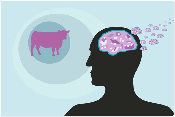
Image Credit: Crystal Eye Studio / Shutterstock.com
The nerve cells are eventually damaged and lost, which causes tiny vacuoles to form in the brain. These give the brain a sponge-like appearance under the microscope, hence the term spongiform disease arose.
This leads to brain damage and the symptoms of prion disease, which include impaired brain function, changes in personality, memory, and behavior, intellectual decline, and movement abnormalities, particularly ataxia. These symptoms usually develop during adulthood and worsen over time, eventually causing death within several years or even a few months.
Prion features
Prions are smaller than viruses and can only be seen through an electron microscope when they have aggregated and formed a cluster. Prions are also unique in that they do not contain nucleic acid, unlike bacteria, fungi, viruses, and other pathogens.
Prions are therefore resistant to procedures that destroy pathogens by breaking down nucleic acid. Furthermore, because these particles are an abnormal version of a normal protein that is already coded for in the body, they do not trigger a host immune response, as other pathogens do.
The normal prion protein is thought to be made up of flexible coils referred to as alpha helices; however, in the abnormally folded form, these helices are stretched out into densely packed structures called beta-sheets. Cellular enzymes referred to as proteases can break down the normal protein; however, prion proteins are unfortunately resistant to this and subsequently accumulate in the brain tissue as they replicate.
Prion-like behavior can also be seen in certain types of fungi. These fungal prions have been studied extensively to provide clues as to how prions affect mammals, although fungal prions are not harmful to their host.
Prion discovery
In the late 1960s, research showed that the agent that causes sheep TSE or scrapie was highly resistant to deactivation by ultraviolet and ionizing radiation, both of which are therapies that would usually destroy any pathogens that contained nucleic acid. However, the nature of these particles was still unclear, which led scientists to make various suggestions including proteins, membrane fragments, small DNA viruses, and polysaccharides.
Some researchers decided that whatever the nature of the agent was, it did not depend on nucleic acid to reproduce. In 1982, Stanley B. Prusiner from the University of California in San Francisco published an article in Science describing the process of purifying the scrapie-causing agent and he described a protein.
Prusiner wrote in the article, "Because the novel properties of the scrapie agent distinguishes it from viruses, plasmids, and viroids, a new term "prion" was proposed to denote a small proteinaceous infectious particle that is resistant to inactivation by most procedures that modify nucleic acids." Prusiner’s discovery led to him being awarded the Nobel Prize in 1997.
References
- https://www.cdc.gov/
- www.nhs.uk/.../Introduction.aspx
- https://www.uclh.nhs.uk/
- http://ghr.nlm.nih.gov/condition/prion-disease
- http://www.bseinfo.org/priondefinition.aspx
Further Reading
Last Updated: Oct 29, 2022