Introduction to the central nervous system (CNS)
The brain
The brainstem
The cerebrum
The cerebellum
The diencephalon
The spinal cord
The meninges
Neurons
The PNS
The somatic nervous system
The autonomic nervous syste
References
Further reading
Introduction to the central nervous system (CNS)
The nervous system is a complex network of nerves and cells that carry messages to and from the brain and spinal cord to various parts of the body. The proper functioning of these nerves ensures that each organ system, such as the cardiovascular, gastrointestinal, and immune systems, can adequately communicate with one another.
The nervous system includes the central nervous system (CNS) and peripheral nervous system (PNS). The CNS is made up of the brain and spinal cord, whereas the PNS is made up of the somatic and autonomic nervous systems.
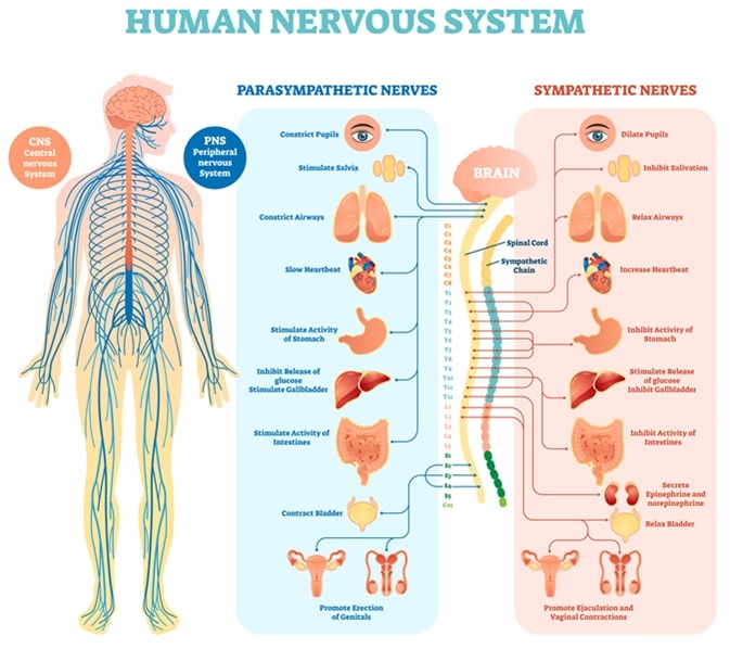
Image Credit: VectorMine / Shutterstock.com
The brain
On average, the brain weighs between 1.3 to 1.4 kg, with about 60% of the brain consisting of fat. The remaining 40% of the brain consists of protein, water, carbohydrates, and salts.
The brain can be divided into four distinct regions: the brainstem, cerebrum, cerebellum, and diencephalon. Taken together, these different areas of the brain control thought, memory, emotion, touch, motor skills, vision, breathing, hunger, temperature, and all other processes that occur within the body.
The brain consists of both gray and white matter. Gray matter, which is darker in color and surrounds white matter, consists of neuron somas, and round central cell bodies. Conversely, white matter, which is lighter in color and comprises the inner portion of the brain, is primarily made up of axons, the long stems that connect neurons.
Gray matter typically processes and interprets information, whereas white matter transmits information to other areas of the nervous system.
Image Credit: ShadeDesign / Shutterstock.com
The brainstem
The brainstem, located in the middle of the brain, is the stalk-like part of the brain that connects the brain to the spinal cord and is only about one inch long. This region regulates essential functions such as blood pressure, breathing, heart rhythms, and swallowing.
The brainstem can be further subdivided into the midbrain, pons, and medulla.
The midbrain, otherwise known as the mesencephalon, is crucial for regulating eye movements, emotions, hearing, and long-term memory. Notably, the substantia nigra, rich in dopamine neurons, is located within the midbrain and is often affected by Parkinson's disease.
The pons is the starting location for four of the 12 cranial nerves. Some of the different functions regulated by the pons include facial movements, hearing, breathing, and balance.
The medulla is located at the bottom of the brainstem where the brain and spinal cord meet. This region of the brainstem regulates breathing, heart rate, and blood pressure. Additionally, the medulla maintains reflective activities such as sneezing, vomiting, coughing, and swallowing.
The cerebrum
The cerebrum is the most significant part of the brain and is lined by a deeply folded layer of nerve tissue called the cerebral cortex. Located at the front of the brain, the cerebrum is divided into the right and left cerebral hemispheres, both connected by the corpus callosum.
The right hemisphere is responsible for creating awareness, emotions, facial expression perception, posture, and prosody, whereas the left hemisphere is dominant in language and pre-processing social emotions. The right hemisphere controls the left side of the body, whereas the left hemisphere controls the right side.
The hemispheres are divided into four lobes, which include the frontal, temporal, parietal, and occipital lobes.
The frontal lobe is anterior to the central sulcus and regulates voluntary movements, speech, memory, emotions, personality, judgment, motor function, planning, organizing, and short-term memory.
The parietal lobe is posterior to the central sulcus and above the occipital lobe. This lobe controls spatial relationships, allowing individuals to understand where their body is compared to surrounding objects. Furthermore, the parietal lobe allows for perceiving sensations like pain and touch.
Broca's and Wernicke's areas are essential for speech production and understanding. Broca's area, which controls the ability to produce speech, is located in the frontal lobe. Conversely, Wernicke's area, which allows individuals to understand spoken language, is located in the parietal lobe.
The temporal lobes are located at the sides of the brain and are inferior to the lateral fissure. These lobes are essential for visual, smell, and taste processing, sound and language interpretation, memory, and hearing.
The occipital lobe is located in the posterior portion of the brain behind the parietal and temporal lobes and is responsible for processing visual data, including colors and shapes.
The cerebellum
The cerebellum is located beneath the temporal and occipital lobes and above the brainstem. The cerebrum is responsible for regulating voluntary motor function, coordination, and balance. Recent studies have indicated that the cerebellum may also be involved in thought, emotions, and social behaviors, as well as the pathophysiology of addiction, autism, and schizophrenia.
The diencephalon
The diencephalon includes the thalamus and hypothalamus. The thalamus is a relay center for sensory data, whereas the hypothalamus transmits hormonal signals to the body through the pituitary gland.
The thalamus and hypothalamus, together with the amygdala and hippocampus, comprise the limbic system. The amygdala regulates emotion, memory, as well as the brain's reward system, stress, and the 'fight or flight' response to threats.
The hippocampus, located underneath each temporal lobe, is vital for long-term memory. This structure also has a role in learning, navigation, and spatial perception.
The spinal cord
The spinal cord is a long tube-like structure that extends from the brain. The spinal cord can be classified into cervical, thoracic, and lumbar areas located in the neck, chest, and lower back.
A total of 31 pairs of nerves and nerve roots comprise the spinal cord. The spinal cord region from which a pair of spinal nerves originates is called the spinal segment.
The cervical spinal cord consists of eight nerves that originate from the neck and run mainly to the face and head.
A total of twelve pairs of nerves can be found within the thoracic region of the spinal cord. These nerves allow for movements in the upper body, including extension of the chest, upper back, and abdomen.
An additional ten pairs of nerves originate from the lower back, with five nerve pairs in the lumbar and sacral regions. The lumbar nerve pairs proceed to the legs and feet, whereas the sacral nerve pairs extend from the low back into the pelvis.
The meninges
The meninges are three membranous layers that cover and protect both the brain and spinal cord. The meninges layers include the dura mater, arachnoid, and pia mater.
The dura mater is the outermost meninges layer and can be further subdivided into the periosteal and meningeal layers. The middle meninges layer is the arachnoid, a web-like layer of connective tissue that does not contain any nerves or blood vessels. Finally, the pia mater is the thinnest meninges layer.
Neurons
The neuron, the basic unit of the nervous system, is a specialized conductor cell that receives and transmits electrochemical nerve impulses between the brain and the rest of the nervous system.
A neuron consists of a cell body, dendrite, and axon. The cell body contains the nucleus, which controls cellular activities and contains genetic material.
Dendrites are branched projections that extend from the cell body and receive signals from other neurons.
Electrical signals travel down a long and thin process known as an axon, which extends from the cell body. These chemical signals, more commonly referred to as neurotransmitters, travel between neurons through a space known as the synapse.
Sensory neurons carry signals from sensory receptors to the brain, whereas motor neurons carry signals from the brain to other nerves, muscles, and glands. The third class of neurons includes interneurons.
The myelin sheath insulates neurons and is continuous along the axons or dendrites, except at the nodes of Ranvier. Myelin, which consists of fat and proteins, provides protection to the neuron, propagates electrical impulses between neurons, and maintains the strength of the signal as it travels down the axon.

Image Credit: MattLphotography / Shutterstock.com
The PNS
The PNS consists of both the somatic and autonomic nervous systems. Taken together, these systems transmit information from different areas of the body to the brain and ensure that signals sent from the brain are transmitted to other areas of the body.
The somatic nervous system
The somatic nervous system (SNS) consists of peripheral nerve fibers that carry sensory information or sensations from peripheral organs to the CNS. The SNS also includes motor nerve fibers that exit the brain to carry commands for movement to the skeletal muscles.
For example, upon touching a hot object, sensory nerves carry information about the heat to the brain. Subsequently, the brain, through motor nerves, commands the hand muscles to withdraw it immediately. This process takes less than one second to complete. The neural cell body that carries this information often lies within the brain or spinal cord and projects directly to a skeletal muscle.
The autonomic nervous system
The autonomic nervous system (ANS) controls the nerves of the body's inner organs that cannot be controlled consciously. The ANS can be further subdivided into the sympathetic, parasympathetic, and enteric nervous systems. Some of the different activities controlled by the ANS include the heartbeat, digestion, subconscious breathing, blood pressure, and sexual arousal.
How Neurotransmission & brain signals work - 3D animation
References
Further Reading
Last Updated: Sep 4, 2022