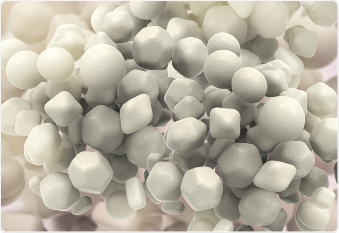
Nanoparticles are 1 – 100 nm in size and can consist of one or a mixture of elements. Nanoparticles are commonly made up of inorganic substances such as gold, silver, or iron oxide, but can also comprise of liposomes and organic structures such as carbon nanotubes.
 Image Credit: Kateryna Kon / Shutterstock
Nanoparticles can be characterized in two ways. The first method of which are methods aiming to determine the physical properties of the nanoparticle itself, such as size, shape, monodispersity, or crystal structure. Other characterization methods aim to determine chemical characteristics of the particle, including the presence and method of bonding of ligands or other conjugated molecules, and the zeta potential.
Physical characterization of nanoparticles
A list of techniques commonly employed to characterize physical aspects of nanoparticles is given below:
- UV-Visible Spectroscopy
- Transmission Electron Microscopy (TEM)
- Dynamic Light Scattering (DLS)
- Differential Centrifugal Sedimentation (DCS)
- Nanoparticle Tracking Analysis (NTA)
- Scanning Electron Microscopy (SEM)
- X-Ray Diffraction (XRD)
Several of these techniques, including DLS, DCS, NTA and TEM focus on determining the dimensions of a particle, and the degree of homogeneity between the particles in a colloid.
A trade-off between good representative data of particles in the whole sample, and more detailed inspection of a single particle exists within these methods, and so several are frequently used in conjunction with one another.
For example, DCS is able to very accurately determine the diameter of millions of spherical particles in a sample to a resolution of less than 1 nm, though results for non-spherical particles will be skewed to indicate that the particles are smaller than they really are.
TEM, on the other hand, can be used to directly photograph clusters of particles on a prepared mesh grid, allowing details of the exact shape of the particle to be imaged. Unfortunately, the number of particles that can be analysed by TEM is unrepresentative of the far larger numbers of particles produced during synthesis.
Nanoparticles made from gold and silver are plasmonic, meaning that they are able to scatter incoming light in such a way as to appear a specific colour. Thanks to this phenomenon, UV-Visible spectroscopy can be used to infer information regarding the shape, size, concentration, and monodispersity of the particles, and even the presence of ligands on the surface of the particle.
Chemical characterization of nanoparticles
Since nanoparticles can be made from a wide variety of materials, chemical characterization methods can vary significantly. Some common techniques are listed below:
- Gel electrophoresis
- 1H/13C nuclear magnetic resonance spectroscopy (NMR)
- Fourier-transform infrared spectroscopy (FTIR)
- Surface enhanced Raman spectroscopy (SERS)
- Biofunctionality Testing (e.g. immunoblotting)
- X-ray photoelectron spectroscopy
- Inductively couple plasma mass spectrometry (ICP-MS)
Techniques such as ICP-MS may be used to determine the concentration of inorganic materials either making up or attached to the nanoparticle, while FTIR and SERS are limited to the determination of molecules attached to the surface of the nanoparticle.
Gel electrophoresis may be used to determine the zeta potential of nanoparticles, and can also act as an indicator of monodispersity or as a functional method of separation of nanoparticles based on size, shape and surface charge.
In a biological setting, assays may be developed to work in conjunction with nanoparticles conjugated with a particular enzyme or protein, with bonding to a complimentary enzyme that is built into the assay able to confirm successful conjugation.
Recently, pregnancy tests incorporating gold nanoparticles have been released to the market, which use plasmonic gold nanoparticles as an indicator.
Further Reading
Last Updated: Nov 2, 2018