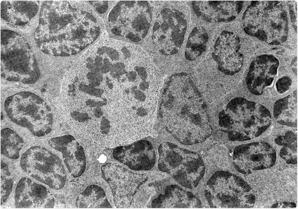
Cryo-electron tomography (CET) is an imaging technology often used for visualizing biomolecules and cells.
 Credit: Jose Luis Calvo/Shutterstock.com
Cryo-electron tomography is a form of transmission electron microscopy, in which a series of two-dimensional images of the sample are captured at different angles in a vacuum environment, and then combined to create a three-dimensional reconstruction.
In cryo-electron tomography, samples are immobilized in ice and imaged at very low temperatures. The sample is dissolved in aqueous media, then plunged into a cryogenic agent to create non-crystalline, or vitreous ice.
The advantage of this approach is avoiding dehydration or chemical fixation that could alter the appearance of structures.
Applications of CET in Microbiology
Compared to other imaging techniques, CET can provide unique details about specimens including subcellular organelles or structurally heterogeneous protein complexes.
Visualizing cytoskeletal networks
Traditional EM for cytoskeletons has provided some insights into the architecture of actin networks, but spatial resolution has been limited. Views of connections between actin filaments are incomplete, and the necessary preparation methods for the sample distorts the structure of the actin networks.
Actin structures are constantly reorganizing in live cells. Vitrification of cells for CET is a key technique for accurate 3D reconstruction of the cytoskeleton.
For example, CET imaging of Dictyostelium discoideum cells showed connections between the actin filament network and the plasma membrane, as well as branching points of large actin filaments.
CET has the potential to increase understanding of specific functional states of cells, such as during particle uptake. The ability to arrest cells instantly can also provide a window into very rapid processes like filopodial protrusion.
Study of infected cells
Some cellular protrusions such as filopodia are prone to viral docking, leading to viral infection. CET can be used to study infected cells, revealing information about the stages of viral assembly and development in the cell.
Tomographic images during viral infection can also provide data on variation between one virus and another, and on changes in distribution between viral strains.
Applications of CET in drug development
Many newer drugs in development have complex formulations designed for improvements in effectiveness and bioavailability. For example, drugs can be encapsulated in a liposome.
These are usually about 100 nm in size, allowing the liposome to move through the bloodstream to its target. CET is a useful tool for studying the drug liposome. Key characteristics of the liposome can be captured in the images, as well as the size and structure of its cargo.
CET as a diagnostic tool
Cilia are external cell structures that play an essential role in human health. In primary ciliary dyskinesia, the cilia have structural defects.
Traditional EM methods have been used to study cilia structures in normal and diseased patients, but due to sample preparation methods required by EM, the disease-related changes in cilia are poorly understood.
Use of CET revealed a previously unknown primary defect and heterogeneous secondary defects in cilia affected by PCD. The data revealed a structural and functional heterogeneity in the radial spokes of the cilia, suggesting a mechanism for PCD, and demonstrated the utility of CET for diagnosing human disease.
Further Reading
Last Updated: Jul 19, 2023