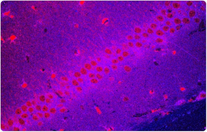
Cytoophidia are filamentous structures present inside a cell. Studies suggest that cytoophidia may have important biological functions in the compartmentalization of cells.
Skip to:
 Oleg Senkov | Shutterstock
In 2010, cytidine triphosphate synthase (CTPS) was independently discovered by three research groups. CTPS is a metabolic enzyme that is responsible for the de novo synthesis of nucleotide cytidine triphosphate (CTP) that can form filamentous structures called cytoophidia. These filamentous structures are found in humans and several other diverse species.
Morphology and function of cytoophidia
Cytoophidia in humans
Cytoophidia are present in several prokaryotes and eukaryotes. However, they differ in number, length, polarity, and localization in across different species. In humans, CTPS is found in both the cytoplasm and nucleus. In the nuclues, cytoophidia are mainly straight, whereas cytoplasmic cytoophidia can be curved, heart, ring-shaped, and curled. Cytoplasmic cytoophidia are longer and have both sharp and blunt ends compared to nuclear cytoophidia. These sturctures are present during all cell cycle stages and can be found within a wide range of organs.
Cytoophidia in Drosophila, bacteria and fungi
The widespread presence of cytoophidia suggests that it may play a role in the regulation of the metabolism. Indeed, it has been discovered within in the optic lobe (the brain structures involved in vision) of Drosophila, where it is essential for homeostasis. It is also present in germline cells where it is thought to be involved in membrane phospholipid synthesis.
In bacteria, CTPS works with bacterial cytoskeletal proteins to regulate the curved morphology of the cell membrane. This function is independent of its enzymatic activity. Similarly, it is present in fungi where it exists in different forms based on the conditions. One of the functions of cytoophidium is to store inactive enzymes that can be released when conditions are favorable. In this way, it is both regulated by, and regulates, metabolism.
Cytoophidium assembly
The cytoophidium is highly sensitive to growth and nutrient conditions. The CTPS filament may be confined to binding sites and its activity may be disrupted in conditions of environmental stress. When the stress is removed, CTPS is again released into the cytoplasm and forms active CTPS.
One of the factors that regulate the assembly and disassembly of cytoophidium is cellular proliferation. For example, the number of cytoophidia in Drosophila neural stem cells increases during the process of differentiation.
Cytoophidia can also be induced in cases where nutrients are limited, including glutamine deprivation. In such cases, the CTPS filament remains inactive. An L-glutamine analog, 6-diazo-5-oxo-L-norleucine (DON), can also promote cytoophidium dissociation. The CTPS filaments formation is inversely related to its ubiquitination and is resistant to the autophagy-lysosome degradation process.
Regulation of cytoophidium assembly
There are several proteins that regulate the assembly and disassembly of CTPS. One such regulator is non-receptor tyrosine kinase DAck. This is the Drosophila homolog of mammalian Ack1. This regulator colocalizes with CTPS in the germline cells of the flies.
Mutations in this protein are related to architecture and morphology aberrations of the CTPS filaments. This often leads to a reduction in fertility. Proto-oncogene Casitas B-lineage lymphoma (Cbl) also controls the structure of cytoophidium.
C-Myc is an oncogene that is upregulated in several cancers. Similarly, CTPS expression is also upregulated in many cancer forms. Studies show that overexpression of Myc can increase the expression of cytoophidium, while the downregulation of Myc can reduce or obstruct cytoophidium formation. Such studies show that CTPS could be a potential therapeutic target in myc-dependent cancers.
Inosine 5[prime]-monophosphate dehydrogenase(IMPDH) has also been shown to upregulate the production of cytoophidium filamentous structures.
Further Reading
Last Updated: May 1, 2019