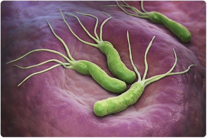
Helicobacter pylori is a Gram-negative, spiral-shaped, flagellated bacterium, that colonizes the gastric mucosa.
Although the gastric mucosa is protected against bacterial infections, H. pylori adapts and resides in the mucus, where it can attach to epithelial cells and colonize the cells within the stomach. Once H. pylori colonizes the stomach, it replicates and releases toxins which can cause diseases such as gastritis, peptic ulcer, or gastric malignancies.

Helicobacter Pylori is a Gram-negative, microaerophilic bacterium found in the stomach. 3D illustration. 3D rendering. Image Credit: Tatiana Shepeleva / Shutterstock
Colonisation and Pathogenesis of Helicobacter pylori
For successful colonization of H. pylori in the stomach, 4 steps are crucial:
- Survival in the acidic stomach.
- Migration towards epithelial mediated by flagella.
- Attachment to host cells by adhesins/receptor interactions.
- Causing tissue damage via toxin release.
H. pylori bacteria contain urea channels that regulate urease activity within the bacteria, these channels control the urea influx to allow the bacteria to survive in the low pH of the stomach.
H. pylori bacteria move towards epithelial cells via the actions of flagella. Many studies have shown that flagella-mediated motility is crucial for the H. pylori colonization of the gastric mucosa.
The interactions between adhesins and receptors allow for the bacteria to attach to cells. Key adhesins that facilitate this process are:
- Blood antigen binding protein A (BabA).
- Sialic acid-binding adhesin (SabA).
- Neutrophil-activating protein (NAP).
- Heat shock protein 60 (Hsp60).
- Adherence-associated proteins (AlpA and AlpB).
Cytotoxin-associated gene A (CagA) protein localizes to the inner surface of the plasma membrane. It undergoes tyrosine phosphorylation, altering the cell signaling within a cell, which alters the adhesion, spreading, and migration of a cell. Vacuolating cytotoxin A (VacA) embeds into the host cell membrane and has the characteristics of an anion-selection channel. The VacA channel allows for the efflux of metabolic substrates from H. pylori for bacterial growth. VacA also induces the release of cytochrome C from mitochondria, ER stress, and apoptosis in host cells.
H Pylori Infection Signs, Symptoms and Treatment
Helicobacter pylori replication
H. pylori can replicate in epithelial cells. It induces the formation of autophagic vesicles, which are the site for its replication. The mechanisms and regulation controlling the growth and proliferation of H. pylori are not well understood. The DNA of H. pylori contains a circular chromosome, but some strains also contain plasmids.
The replication of DNA in H. pylori occurs in 3 steps:
- Initiation
- Elongation
- Termination
Chromosome replication starts at a site called the oriC site. The assembly of two replisomes establishes two replication forks in opposite orientations. The replication forks proceed bi-directionally until they meet at the opposite side of the chromosome (the terminus region). Initiation of DNA replication takes place when the action of various proteins results in the formation of a ‘bubble’ near the oriC site. This allows for the unwinding of the DNA and short RNA primers are made on the leading strand. DNA polymerase III can start to extend the DNA replication from the RNA primer. DNA ligase allows for the joining of the replicated strands. The lagging strand is synthesized by the discontinuous synthesis of small ‘Okazaki fragments’.
In H. pylori, the termination of DNA replication and chromosome segregation are not the same that is seen in other bacteria such as E. coli. The mechanisms of these processes are not well known.
In summary, H. pylori is well adapted to survive in the highly inhabitable environment of the stomach. These adaptations allow for the directed migration of the bacteria towards target cells and colonization of the bacteria. Following colonization, the bacteria can replicate and begin to release toxins. This will eventually lead to the development of diseases in a host.
More research into the life cycle of H. pylori will allow for a better understanding of each process that has been previously discussed. This will provide better tools to treat the diseases caused by H. pylori.
Further Reading
Last Updated: Feb 26, 2019