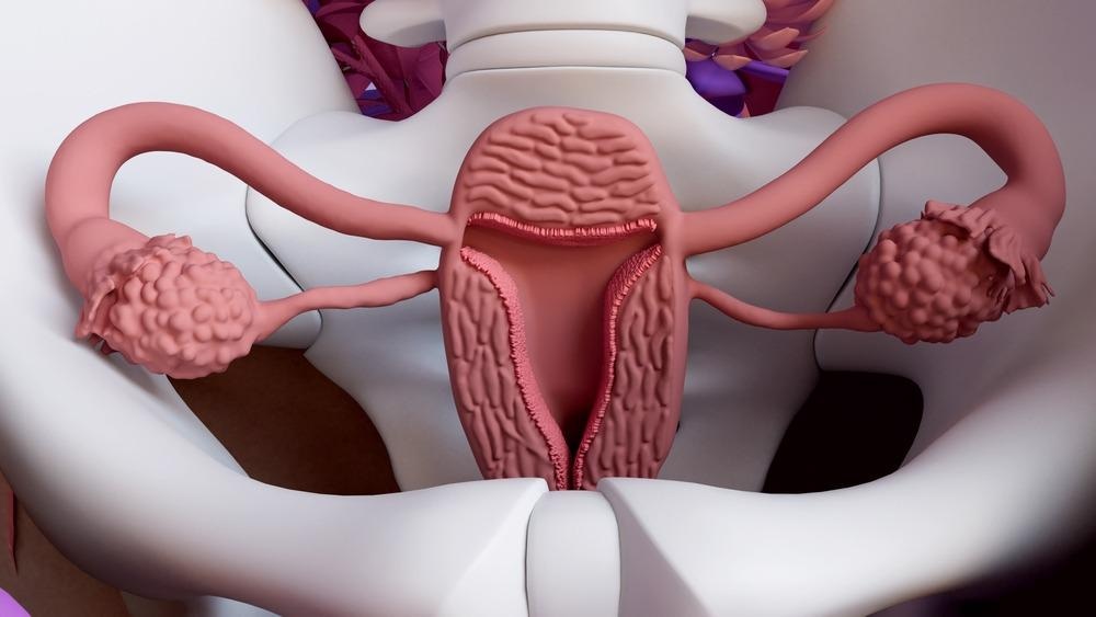
The uterus is the organ that hosts a developing fetus during pregnancy. This article will look at the uterus’s biochemistry and explore its biochemical and physiological changes as pregnancy progresses to full term.

Image Credit: Design_Cells/Shutterstock.com
The Uterus
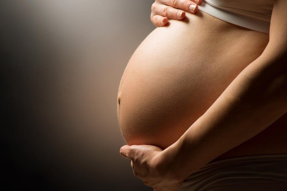
The uterus is part of the female reproductive system, responsible for the healthy embryo and fetal development and it undergoes several changes during pregnancy. This organ is distensible and can expand to a size that can accommodate a fully-grown uterus. During birth, the uterus pushes the baby through the vagina using strong muscles. After birth, the uterus will usually return to its original size at around the six-week mark. Normal human pregnancy lasts around 40 weeks.
Structure of the Uterus
The uterus is located in the pelvic region of the body, behind the bladder (and almost overlying it), and in front of the sigmoid colon. Anatomically, it is divided into four regions – the fundus, corpus, cervix, and cervical canal. The cervix protrudes into the vagina, and the uterus is held in place in the pelvis by several ligaments, including the cardinal ligaments, the pubocervical ligaments, and the uterosacral ligaments.
The uterine wall consists of three layers, the perimetrium, myometrium, and the endometrium. The endometrium, which is the innermost layer, has a mucus membrane. Uterine glands and blood vessels in the endometrium increase in size and number, forming the decidua. The myometrium is mainly composed of smooth muscle. The perimetrium, the outermost layer, is a layer of the visceral peritoneum.
A layer of fatty and fibrous connective tissue called the parametrium covers the outside surface. This connects the uterine tissue to the other pelvic tissues. The uterine microbiome consists of commensal organisms present in the uterus.
Changes Inside the Uterus
Aside from size changes, there are many changes to the uterus during pregnancy. The corpus luteum, the structure that envelops the embryo, develops soon after fertilization. The corpus luteum releases pregnancy hormones including progesterone. Progesterone stops the uterus from contracting normally as it would during menstruation, as well as controlling the growth of the uterine wall lining.
Around days 6-12 after fertilization, tiny, finger-like projections start to develop which grow to form the placenta, the structure responsible for providing nutrients to the developing embryo. Progesterone and estrogen secreted by the placenta also encourage further physiological changes in the embryo.
After the four-week mark, blood vessels grow and enlarge to support the development of the embryo. Additional changes are the softening of the cervix, the development of a mucus plug, changes in myometrium muscles, and ligament changes. After birth, the placenta is pushed out of the vagina along with the baby.
The Placenta
The placenta is a unique structure that is only formed during pregnancy. It is composed of cells that originate from the blastocyst. Cytotrophoblasts and syncytiotrophoblasts line the placenta and breach the uterine wall to remodel the blood vessels contained in the uterus, providing oxygen and nutrients for the developing fetus and removing waste. The placenta helps to protect the developing fetus from disease.
Many hormones are found in the placenta, including progesterone, estradiol, estriol, chorionic gonadotropin, and human placental lactogen. These hormones support the healthy growth of the fetus during pregnancy in multiple ways. The placenta also contains other proteins such as alpha protein, inhibin A, and pregnancy-associated plasma protein-A.

Image Credit: Subbotina Anna/Shutterstock.com
Changes in Maternal Uterine Fluid
The biochemical changes in the uterus during pregnancy are highly complex. The exact metabolic and biochemical changes in the maternal uterine fluid are still not properly understood. Several changes in metabolism changes occur up to 1-4 of pregnancy that has been linked to the development of the blastocyst and protecting it from the environment in the uterus.
Changes occur during early implantation in the levels of acids such as palmitoleic acid and fumaric acid, suggesting a link between the presence of these chemicals and endometrial receptivity. Changes in metabolism including the downregulation of aerobic respiration and the upregulation of glyoxylic metabolism have occurred during early implantation in both maternal and uterine plasma.
It has been suggested that biochemical and metabolic changes in the maternal fluid are involved with several important processes in fetal development including energy preservation, utilization of alternative nutrients, and helping regulate the interface between mother and fetus.
Summing Up
The uterus is a complex organism that undergoes biochemical and physiological changes during pregnancy. Understanding how the uterus functions and changes during pregnancy is key to ensuring a healthy birth and diagnosing and treating diseases and conditions that otherwise may cause problems during pregnancy for both the baby and the mother. Whilst some aspects of the biochemical composition of the uterine environment are poorly understood, studies are revealing more information about this organ.
References:
Further Reading
Last Updated: Apr 13, 2022