The current state of the reagents market

Image Credit: Caleb Foster/Shutterstock.com
The types of imaging reagents utilized by the biological and medical imaging reagents market include fluorescent probes and dyes, fluorescent proteins, nanoparticles, magnetic resonance imaging, ultrasound and X-ray contrast reagents, and imaging radiopharmaceuticals.
The biotechnology, life sciences, and medical and pharmaceutical industries rely heavily on imaging reagents, with the field of diagnostics being the largest market for imaging reagent products.
In the last few years research has been helping to advance the use of reagents and their associated technologies to develop new and better diagnostic applications. A current main driver in this part of the industry is to establish early detection techniques for Alzheimer's disease and various cancers.
A new and rapidly growing segment of the industry is that of imaging reagents that utilize semiconductor dots, while huge advancements have recently been made in the use of carbon nanomaterials in nuclear imaging techniques. Currently, there are numerous other types of nanoparticle being investigated to assess their potential use in imaging reagents, which will see nanoparticles become a more established segment of the imaging reagents market.
Below we discuss the different types of imaging reagents.
Fluorescent probes and dyes
Many types of fluorescent compounds are chelating agents with humic characteristics, meaning that they react with metal ions to form a stable, water-soluble complex, and they bind to positively charged metal cations and other compounds.
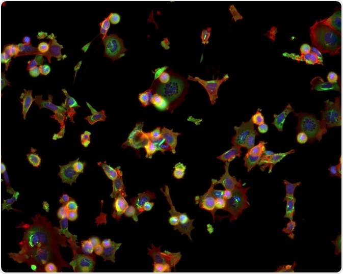
Fluorescent probes are one of the most common reagents used for classifying targeted molecules. They are used to label antibodies of interest, allowing for their detection. They have applications in detecting the location of targeted proteins, as well as in identifying their activation.
They can be used to distinguish protein complex formation and conformational changes, as well as monitor processes in vivo. As time goes on, their versatility, sensitivity, and quantitative capabilities continue to grow, resulting in their increasing popularity.
Fluorescent proteins
Fluorescent proteins are primarily used in gene expression studies to track the localization and dynamics of proteins, organelles, and other cellular components. They are also used as a tracer of intracellular protein trafficking. Techniques such as confocal, wide-field, and multiphoton microscopy are used to view the fluorescent proteins and reveal data on cell structure and function.
Nanoparticles
Nanoparticles have become a focus of many areas of science. Various sectors are exploring how these particles, with their unique characteristics, can be exploited in research and in enhancing and developing numerous applications. Given that nanoparticles are just 1 – 100 nm in diameter, they are a comparable size to biological units.
Nanoparticles are a current focus of cancer research, with scientists aiming to capitalize on the unique properties of the particles, such as their tunable absorption and emission properties, diverse surface chemistries, and unique magnetic properties, which may see them being used as probes for early detection for various kinds of cancer and other diseases. However, nanoparticles themselves pose potential toxicity due to the materials used to construct them. They also have the potential to interfere with other medical tests, so there is a lot of research and development ahead for the use of this kind of imaging reagent before they see widespread use.
Magnetic resonance imaging
Magnetic resonance imaging (MRI) relies on the magnetic properties of materials to generate cross-sectional images of structures within the body. Reagents are used to enhance the visibility of the structures that are picked up on by MRI, and these reagents are known as contrast reagents. Typically, most agents are gadolinium-based, and in-fact, only gadolinium chelated contrast agents are considered safe for human use.
Ultrasound and X-ray contrast reagents
To enhance the visibility of structures identified by X-ray techniques, such as computed tomography, projection radiography, and fluoroscopy, radiocontrast agents are used. Iodine is the most common substance used for these agents, but sometimes barium-sulfate may be used.
Ultrasound also requires specific reagents to enhance the images produced. The agents used in this method are essentially microscopic bubbles of gas that are contained within a thin flexible shell. Different contrast agents use different gases and shell materials.
However, the size of the microbubbles generally measures between 1 and 4 micrometers in size, making them smaller than red blood cells, so easy to circulate through the blood vessels without the risk of them diffusing out. They are then detected when the ultrasound probe emits high-frequency sound waves that hit the agents, causing them to oscillate and reflect a non-characteristic echo.
Imaging radiopharmaceuticals
Radiopharmaceuticals are reagents used by single-photon emission computed tomography (SPECT) and positron emission tomography (PET). These are two of the most highly used and advanced imaging technologies that are currently available.
They are essential to the diagnosis and monitoring of cardiovascular, oncological, neurological, and numerous other types of disease. The most commonly used imaging reagents are forms of fluorine, gallium, and nitrogen, for use with PET, and galium, indium, iodine, technetium, and xenon for use with SPECT.
Summary
There are numerous kinds of imaging reagents, each of which have been developed with specific imaging techniques in mind. The future will likely see major development in the use of nanoparticles as imaging reagents, as researchers discover more about their potential functions.
Sources:
- Biologic and Medical Imaging Reagents: Global Markets. (2017). [online] BCC Research. Available at: https://www.reportlinker.com/p05142314/Biologic-and-Medical-Imaging-Reagents-Global-Markets.html [Accessed 27 Nov. 2019].
- Choi, H. and Frangioni, J. (2010). Nanoparticles for Biomedical Imaging: Fundamentals of Clinical Translation. Molecular Imaging, 9(6), pp.7290.2010.00031. https://www.ncbi.nlm.nih.gov/pmc/articles/PMC3017480/
- Hermanson, G. (2013). Fluorescent Probes. Bioconjugate Techniques, pp.395-463. https://www.sciencedirect.com/science/article/pii/B9780123822390000108
- Lusic, H. and Grinstaff, M. (2012). X-ray-Computed Tomography Contrast Agents. Chemical Reviews, 113(3), pp.1641-1666. https://www.ncbi.nlm.nih.gov/pmc/articles/PMC3878741/
- Nune, S., Gunda, P., Thallapally, P., Lin, Y., Laird Forrest, M. and Berkland, C. (2009). Nanoparticles for biomedical imaging. Expert Opinion on Drug Delivery, 6(11), pp.1175-1194. https://www.ncbi.nlm.nih.gov/pmc/articles/PMC3097035/
Further Reading
Last Updated: Feb 6, 2020